Reviewed by — Neurosurgeon · Sidra Medicine, Doha Last updated:

Overview

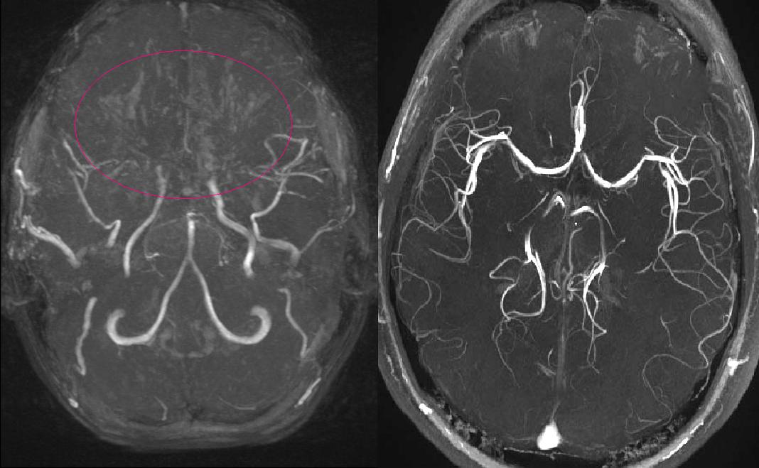
Trauma is the leading cause of death in children beyond infancy, and traumatic brain injury (TBI) is the dominant mechanism. Paediatric TBI differs from adult TBI in mechanism, anatomy, physiology, and outcome, and is managed according to dedicated paediatric guidelines (Brain Trauma Foundation, 3rd Edition, 2019). Cerebrovascular disease in children, though far less common than in adults, includes several conditions essentially unique to childhood: vein of Galen aneurysmal malformation, paediatric moyamoya disease and syndrome, and stroke patterns specific to sickle-cell disease.

Pediatric severe TBI management is now systematised by the BTF 3rd Edition guidelines (Kochanek et al., 2019). Triage of minor head injury is informed by the PECARN clinical decision rules (Kuppermann et al., 2009). Cerebral AVM risk stratification continues to rely on the Spetzler-Martin grade (Spetzler & Martin, 1986), supplemented by supplementary scoring systems in selected centres.

References used here

  1. Kochanek PM, Tasker RC, Carney N, Totten AM, Adelson PD, Selden NR, Davis-O'Reilly C, Hart EL, Bell MJ, Bratton SL, Grant GA, Kissoon N, Reuter-Rice KE, Vavilala MS, Wainwright MS. Guidelines for the Management of Pediatric Severe Traumatic Brain Injury, Third Edition: Update of the Brain Trauma Foundation Guidelines. Pediatr Crit Care Med. 2019;20(3S Suppl 1):S1-S82.
  2. Kuppermann N, Holmes JF, Dayan PS, Hoyle JD Jr, Atabaki SM, Holubkov R, Nadel FM, Monroe D, Stanley RM, Borgialli DA, Badawy MK, Schunk JE, Quayle KS, Mahajan P, Lichenstein R, Lillis KA, Tunik MG, Jacobs ES, Callahan JM, Gorelick MH, Glass TF, Lee LK, Bachman MC, Cooper A, Powell EC, Gerardi MJ, Melville KA, Muizelaar JP, Wisner DH, Zuspan SJ, Dean JM, Wootton-Gorges SL; Pediatric Emergency Care Applied Research Network (PECARN). Identification of children at very low risk of clinically-important brain injuries after head trauma: a prospective cohort study. Lancet. 2009;374(9696):1160-1170.
  3. Scott RM, Smith ER. Moyamoya disease and moyamoya syndrome. N Engl J Med. 2009;360(12):1226-1237.
  4. Spetzler RF, Martin NA. A proposed grading system for arteriovenous malformations. J Neurosurg. 1986;65(4):476-483.
  5. Albright AL, Pollack IF, Adelson PD. Principles and Practice of Pediatric Neurosurgery. 3rd Edition. Thieme, 2015. ISBN: 978-1-60406-799-6.
  6. Winn HR (Editor). Youmans and Winn Neurological Surgery. 8th Edition (4-volume set). Elsevier, 2022. ISBN: 978-0-323-66192-8.
Trauma

Pediatric Traumatic Brain Injury; Overview

WHO 2021 classification: Acute injury to the brain caused by external mechanical force. Severity is graded by post-resuscitation Glasgow Coma Scale (GCS): mild (GCS 13-15), moderate (GCS 9-12), and severe (GCS ≤8). The Pediatric GCS modifies the verbal subscale for children younger than 5 years.

TBI is the leading cause of death and acquired disability in children beyond infancy. Mechanisms differ by age; falls and abusive head trauma dominate in infancy; sport, recreational and pedestrian injuries in school-age children; motor-vehicle collisions in adolescents. The neurosurgeon's task is two-fold: prevent secondary injury and address surgical mass lesions.

Epidemiology

Incidence
Paediatric TBI accounts for hundreds of thousands of emergency department visits annually in high-income countries; severe TBI (GCS ≤8) is a minority but accounts for the bulk of mortality and long-term disability.
Age peak
Bimodal: infants/toddlers (falls, abusive head trauma) and adolescents (sport, road traffic).
Location
Brain; diffuse and focal injury patterns coexist; extra-axial collections (epidural, subdural), contusions, and diffuse axonal injury are the major patterns.

Clinical Presentation

  • Initial assessment by the ATLS/APLS framework: airway with cervical-spine protection, breathing, circulation, disability (GCS, pupils), exposure.
  • Mild TBI (GCS 13-15): the great majority of presentations; applies PECARN-type clinical decision rules to limit unnecessary CT.
  • Moderate (GCS 9-12) and severe (GCS ≤8) TBI: ICU-level management, urgent neurosurgical assessment, prevention of secondary injury.
  • Post-concussion symptoms (headache, sleep disturbance, mood change, cognitive complaints): often under-recognised in children; activity-modification and graded return are central to recovery.

Imaging

  • CT is the first-line imaging in the acute setting; fast, available, identifies the surgical lesions (epidural, subdural, contusion, mass effect, midline shift).
  • MRI for sub-acute imaging; superior for diffuse axonal injury (DAI), small contusions, brainstem injury, and follow-up.
  • Skull radiographs have a limited role and are not a substitute for CT in suspected significant injury.
  • Cervical-spine imaging is integral whenever indicated; TBI patients are spinal-injury patients until proven otherwise.

Pathology & Molecular

Histology. Diffuse axonal injury (DAI): axonal shearing at grey-white interfaces, corpus callosum, and brainstem. Focal contusion: cortical haemorrhagic injury, classically at coup and contrecoup sites.

Molecular. Secondary-injury cascades involve excitotoxicity, oxidative stress, inflammation, and apoptosis, the basis of current and trial-stage neuroprotective strategies.

Management

Surgery. Surgical management of identified lesions: evacuation of epidural and subdural haematomas with significant mass effect; elevation of depressed skull fractures meeting criteria; decompressive craniectomy in selected refractory cases.

Adjuvant therapy. Non-surgical critical-care management: stepped algorithm per BTF 3rd Edition; head elevation, normothermia, normocapnia, sedation/analgesia, osmotherapy (hypertonic saline preferred in paediatrics), CSF drainage, paralysis, and consideration of decompressive craniectomy or barbiturate coma for refractory intracranial hypertension.

Considerations. Always image and protect the cervical spine; always exclude non-accidental injury in young children with unexplained TBI; involve rehabilitation services early.

Outcomes

Outcomes correlate strongly with initial GCS, pupillary response, and CT findings. Children generally have better functional outcomes than adults with comparable injury severity, but the long-term neurocognitive impact (especially on attention, executive function, and academic progress) is substantial and often under-recognised.

Clinical Pearls

  • A 'good' GCS does not exclude significant injury; use PECARN-style decision rules and a low threshold for repeat assessment.
  • Cervical spine first; any TBI patient is a cervical-spine patient until imaging and exam clear it.
  • Always ask: could this be non-accidental?, particularly in infants with disproportionate injury.

Glasgow Coma Scale; adult and paediatric (verbal subscale)

CategoryScoreAdult / Child >5yChild <5y
Eye opening (E)4 / 3 / 2 / 1Spontaneous / To voice / To pain / NoneSame as adult
Verbal (V)5OrientedCoos, babbles
Verbal (V)4ConfusedIrritable cries
Verbal (V)3Inappropriate wordsCries to pain
Verbal (V)2Incomprehensible soundsMoans to pain
Verbal (V)1NoneNone
Motor (M)6 / 5 / 4 / 3 / 2 / 1Obeys / Localises / Withdraws / Abnormal flexion / Extension / NoneSame as adult (assessed by observation in infants)
Total GCS ranges 3-15. Severity: severe ≤8; moderate 9-12; mild 13-15. The paediatric verbal subscale is modified for pre-verbal and early-verbal children.

TBI severity classification (by post-resuscitation GCS) and broad management implications

SeverityPost-resuscitation GCSTypical imagingDisposition / management
Mild13-15Selective CT (PECARN-guided)ED observation, discharge with safety advice, follow-up for post-concussion symptoms
Moderate9-12CT (often serial)Admission, neurosurgical consultation, ICU consideration
Severe≤8 (or rapid deterioration)CT, MRI sub-acuteICU admission, intubation, BTF guideline-based management, neurosurgical lesion management
Adapted from the Brain Trauma Foundation Pediatric Severe TBI Guidelines (Kochanek et al., 2019).

References used here

  1. Kochanek PM, Tasker RC, Carney N, Totten AM, Adelson PD, Selden NR, Davis-O'Reilly C, Hart EL, Bell MJ, Bratton SL, Grant GA, Kissoon N, Reuter-Rice KE, Vavilala MS, Wainwright MS. Guidelines for the Management of Pediatric Severe Traumatic Brain Injury, Third Edition: Update of the Brain Trauma Foundation Guidelines. Pediatr Crit Care Med. 2019;20(3S Suppl 1):S1-S82.
  2. Kuppermann N, Holmes JF, Dayan PS, Hoyle JD Jr, Atabaki SM, Holubkov R, Nadel FM, Monroe D, Stanley RM, Borgialli DA, Badawy MK, Schunk JE, Quayle KS, Mahajan P, Lichenstein R, Lillis KA, Tunik MG, Jacobs ES, Callahan JM, Gorelick MH, Glass TF, Lee LK, Bachman MC, Cooper A, Powell EC, Gerardi MJ, Melville KA, Muizelaar JP, Wisner DH, Zuspan SJ, Dean JM, Wootton-Gorges SL; Pediatric Emergency Care Applied Research Network (PECARN). Identification of children at very low risk of clinically-important brain injuries after head trauma: a prospective cohort study. Lancet. 2009;374(9696):1160-1170.
  3. Albright AL, Pollack IF, Adelson PD. Principles and Practice of Pediatric Neurosurgery. 3rd Edition. Thieme, 2015. ISBN: 978-1-60406-799-6.
  4. Winn HR (Editor). Youmans and Winn Neurological Surgery. 8th Edition (4-volume set). Elsevier, 2022. ISBN: 978-0-323-66192-8.
  5. Greenberg MS. Greenberg's Handbook of Neurosurgery. 10th Edition. Thieme, 2023. ISBN: 978-1-68420-504-2.
ICU / Surgical

Severe Pediatric TBI Management

WHO 2021 classification: Children with post-resuscitation GCS ≤8 (or rapid deterioration) require structured intensive care according to the Brain Trauma Foundation Pediatric Severe TBI Guidelines, 3rd Edition (Kochanek et al., 2019). The framework is a stepped algorithm of escalating interventions to control intracranial pressure (ICP) and maintain adequate cerebral perfusion.

The management of severe paediatric TBI is one of the most protocolised areas of neurosurgical ICU care. Outcomes have improved over decades through better adherence to evidence-informed protocols rather than any single breakthrough therapy.

Epidemiology

Incidence
A minority of paediatric TBI but accounts for the great majority of mortality and severe disability.
Age peak
Bimodal; infants (often abusive head trauma) and adolescents (road traffic, sport).
Location
Brain; frequently with associated extracranial injury in road-traffic and high-energy mechanisms.

Clinical Presentation

  • Initial assessment by ATLS/APLS principles with full cervical-spine protection.
  • Intubation for GCS ≤8 (or deterioration); avoid hypoxia and hypotension, both independently worsen outcome.
  • Urgent CT to identify surgical lesions; ICP monitor consideration once stabilised.
  • Continuous physiological targets: oxygenation, CO2 control, cerebral perfusion pressure (CPP), temperature, glucose.

Imaging

  • Initial CT for the surgical decision (evacuate vs medical management) and to define baseline.
  • Repeat CT for clinical deterioration or scheduled re-assessment in the first 24-48 hours.
  • MRI in the sub-acute period for DAI, brainstem injury, and prognostication.

Pathology & Molecular

Histology. Mixed primary injury (contusions, DAI, extra-axial collections) plus secondary injury (oedema, ischaemia, oxidative injury).

Molecular. Secondary-injury cascades (excitotoxicity, oxidative stress, inflammation) drive much of the modifiable injury.

Management

Surgery. Evacuation of mass-effect-producing extra-axial haematoma (epidural or subdural) is urgent. Depressed skull fracture meeting criteria is elevated. Decompressive craniectomy may be undertaken for refractory raised ICP not controlled by tier-1 and tier-2 medical measures.

Adjuvant therapy. Tiered medical therapy per BTF 3rd Edition: tier 1, head-up positioning, normothermia, normocapnia (PaCO2 35-38 mm Hg), adequate sedation/analgesia, osmotherapy (hypertonic saline preferred in paediatrics; mannitol acceptable), CSF drainage via EVD; tier 2, neuromuscular blockade, mild hypocapnia (still within safe limits), barbiturate trial; tier 3; decompressive craniectomy or moderate-to-deep hypothermia (the latter not first-line and used with caution).

Considerations. Avoid prolonged or aggressive hyperventilation. Avoid prophylactic hypothermia. Glucocorticoids are not recommended.

Outcomes

Mortality from severe paediatric TBI has fallen with modern guideline-based care but remains substantial. Long-term cognitive, behavioural, and academic outcomes are determined by injury severity, secondary insults, and rehabilitation.

Clinical Pearls

  • Avoid hypoxia (SpO2 <90%) and hypotension, both independently increase mortality.
  • Hypertonic saline is the preferred paediatric osmotherapy in current BTF guidelines.
  • Hyperventilation has a limited place; use briefly for impending herniation, not as routine therapy.

Stepped management of refractory ICP in paediatric severe TBI (BTF 3rd Edition, simplified)

TierInterventionNotes
FoundationalHead-up 30°, normothermia, normocapnia, adequate sedation/analgesia, normal Na+ and glucoseApply continuously from the outset
Tier 1Osmotherapy: hypertonic saline (3% or higher); mannitol as alternativeHypertonic saline preferred in paediatrics; monitor sodium and osmolality
Tier 1CSF drainage via EVDEffective and reversible; consider early if monitor placed
Tier 2Neuromuscular blockade with continued analgesia/sedationLoss of clinical examination; relies on monitoring
Tier 2Controlled mild hypocapnia (PaCO2 30-35 mm Hg)Avoid prolonged or aggressive hyperventilation
Tier 2Barbiturate trial (e.g., thiopental, pentobarbital)Requires invasive monitoring and haemodynamic support
Tier 3Decompressive craniectomyConsidered in refractory cases; timing and patient selection critical
Tier 3Moderate hypothermia (32-33 °C)Not first-line; risks must be balanced against benefit
Adapted from Kochanek et al., Guidelines for the Management of Pediatric Severe TBI, 3rd Edition. Pediatr Crit Care Med 2019;20(3S):S1-S82. See References.

References used here

  1. Kochanek PM, Tasker RC, Carney N, Totten AM, Adelson PD, Selden NR, Davis-O'Reilly C, Hart EL, Bell MJ, Bratton SL, Grant GA, Kissoon N, Reuter-Rice KE, Vavilala MS, Wainwright MS. Guidelines for the Management of Pediatric Severe Traumatic Brain Injury, Third Edition: Update of the Brain Trauma Foundation Guidelines. Pediatr Crit Care Med. 2019;20(3S Suppl 1):S1-S82.
  2. Albright AL, Pollack IF, Adelson PD. Principles and Practice of Pediatric Neurosurgery. 3rd Edition. Thieme, 2015. ISBN: 978-1-60406-799-6.
  3. Winn HR (Editor). Youmans and Winn Neurological Surgery. 8th Edition (4-volume set). Elsevier, 2022. ISBN: 978-0-323-66192-8.
Trauma; non-accidental

Abusive Head Trauma (AHT)

WHO 2021 classification: Inflicted head injury in infants and young children, most often resulting from violent shaking, blunt-force impact, or both. The term 'abusive head trauma' is endorsed by the AAP in preference to historical terms (e.g., 'shaken baby syndrome') that imply a specific mechanism.

Abusive head trauma is among the most consequential diagnoses in paediatric neurosurgery, the leading cause of TBI death in infants under one year in many series, and a diagnosis with profound medical, child-protection, and medico-legal implications. Multidisciplinary assessment is mandatory.

Epidemiology

Incidence
Estimated 20-30 per 100,000 in infants under 1 year in published series; the true incidence is almost certainly higher (under-recognition is well-documented).
Age peak
Infants; peak incidence in the first 6 months of life; rare after age 4-5.
Location
Brain (subdural haematoma, parenchymal injury), retina (haemorrhages), skeletal (rib, metaphyseal, healing fractures), skin.

Clinical Presentation

  • Presentation may be subtle (irritability, lethargy, vomiting, apnoea, seizures, encephalopathy) and is often disproportionate to the history offered.
  • Inconsistent, changing, or absent history of trauma is itself a red flag.
  • Always examine for retinal haemorrhages (dilated fundoscopy by ophthalmology), cutaneous bruising in young pre-mobile infants, and skeletal injury (skeletal survey).

Imaging

  • CT acutely for surgical decision-making.
  • MRI of brain and (when indicated) cervical spine for full characterisation; sub-acute and chronic blood, parenchymal injury, ligamentous injury at the cervico-medullary junction.
  • Skeletal survey (radiographic) per AAP protocol, with selective bone scintigraphy if survey is inconclusive.

Pathology & Molecular

Histology. Subdural haematoma (often bilateral and of different ages), parenchymal injury, hypoxic-ischaemic injury, retinal haemorrhages.

Molecular. Not applicable; clinical diagnosis.

Management

Surgery. Evacuation of significant subdural collections producing mass effect; treatment of associated injuries; CSF diversion if hydrocephalus develops in the recovery phase.

Adjuvant therapy. Critical-care management for severe cases per BTF guidelines.

Considerations. Mandatory child-protection notification per local jurisdiction. Multidisciplinary team: paediatric neurosurgery, paediatrics, child-protection (safeguarding), ophthalmology, social work, radiology. Documentation must be meticulous, with the diagnosis stated clearly in clinical notes.

Outcomes

Substantial mortality (commonly cited 15-25%) and high rate of severe long-term disability among survivors; neurodevelopmental, motor, visual, and behavioural. Recurrence of abuse is a recognised risk if the diagnosis is missed.

Clinical Pearls

  • If the history does not fit the injury, take it seriously, under-diagnosis of AHT carries the risk of fatal recurrence.
  • Retinal haemorrhages and unexplained skeletal injuries in young infants demand a full work-up, not reassurance.
  • Use the term 'abusive head trauma' in clinical documentation per the AAP statement (Christian & Block, 2009).

References used here

  1. Christian CW, Block R; Committee on Child Abuse and Neglect, American Academy of Pediatrics. Abusive head trauma in infants and children. Pediatrics. 2009;123(5):1409-1411.
  2. Albright AL, Pollack IF, Adelson PD. Principles and Practice of Pediatric Neurosurgery. 3rd Edition. Thieme, 2015. ISBN: 978-1-60406-799-6.
  3. Winn HR (Editor). Youmans and Winn Neurological Surgery. 8th Edition (4-volume set). Elsevier, 2022. ISBN: 978-0-323-66192-8.
  4. Greenberg MS. Greenberg's Handbook of Neurosurgery. 10th Edition. Thieme, 2023. ISBN: 978-1-68420-504-2.
Trauma

Pediatric Skull Fractures

WHO 2021 classification: Fractures of the skull in children. Classified as linear (the great majority), depressed (with bony fragment displaced inward), basal (involving the skull base), and growing (a paediatric-specific late complication of a fracture with underlying dural tear and brain pulsation).

Most paediatric skull fractures are simple, linear, isolated, and managed conservatively. The neurosurgeon's role is to identify the small subset requiring intervention (depressed fractures meeting criteria, basal fractures with CSF leak or cranial-nerve injury, growing fractures) and to recognise the relationship between fracture and underlying brain injury.

Epidemiology

Incidence
Common, a frequent finding on CT performed for paediatric head injury.
Age peak
Throughout childhood; growing skull fractures particularly in infants under 3 years owing to the dynamic skull-brain relationship.
Location
Calvarial (frontal, parietal, occipital) most common; basal in higher-energy injuries.

Clinical Presentation

  • Linear fracture: often clinically silent; identified on CT or as a step palpable beneath the scalp; conservative management is usual.
  • Depressed fracture: palpable depression; consider operative elevation if depression > thickness of skull, associated with dural tear, parenchymal injury, cosmetic concern, or open injury.
  • Basal skull fracture: signs include 'raccoon eyes' (periorbital ecchymosis), Battle's sign (mastoid ecchymosis), haemotympanum, CSF rhinorrhoea or otorrhoea, and cranial-nerve deficits (especially CN I and VII).
  • Growing skull fracture: an enlarging, pulsatile scalp swelling weeks to months after a known fracture in an infant; characteristic and requires surgical repair.

Imaging

  • CT is the workhorse; bony detail, identification of depressed fractures, and assessment of underlying brain injury.
  • MRI for suspected dural tear (growing skull fracture risk), brain injury characterisation.
  • Plain skull radiographs have a limited role and are not a substitute for CT in significant injury.

Pathology & Molecular

Histology. Not a histological diagnosis.

Molecular. Not applicable.

Management

Surgery. Linear isolated fracture: conservative, with imaging follow-up if growing fracture risk. Depressed fracture meeting criteria (depression > skull thickness, open injury, dural tear, mass effect, cosmetic concern in visible area): elevation, dural repair if torn, fragment replacement or cranioplasty. Growing skull fracture: surgical repair of dural defect with cranial reconstruction; early repair prevents progressive enlargement and underlying brain herniation. Basal fracture with CSF leak: most resolve with conservative management; persistent or recurrent leak warrants imaging and consideration of repair (endoscopic endonasal or transcranial depending on location).

Adjuvant therapy. Antibiotic prophylaxis for open or depressed fractures per institutional protocol; vaccination status review.

Considerations. An infant with a linear fracture and underlying dural tear is at risk of growing skull fracture; schedule follow-up imaging at 1-3 months.

Outcomes

Excellent for isolated linear fractures. Outcomes for depressed fractures depend principally on the associated brain injury. Growing skull fracture has a good outcome when recognised and repaired early.

Clinical Pearls

  • Most paediatric skull fractures are linear and need no surgery, the neurosurgeon's job is to identify the few that do.
  • An enlarging, pulsatile scalp swelling in an infant weeks after a head injury is a growing skull fracture until proven otherwise.
  • A basal skull fracture with CSF rhinorrhoea usually resolves with conservative management; do not rush to surgery.

References used here

  1. Albright AL, Pollack IF, Adelson PD. Principles and Practice of Pediatric Neurosurgery. 3rd Edition. Thieme, 2015. ISBN: 978-1-60406-799-6.
  2. Winn HR (Editor). Youmans and Winn Neurological Surgery. 8th Edition (4-volume set). Elsevier, 2022. ISBN: 978-0-323-66192-8.
  3. Greenberg MS. Greenberg's Handbook of Neurosurgery. 10th Edition. Thieme, 2023. ISBN: 978-1-68420-504-2.
Trauma; spine

Pediatric Cervical Spine Injury & SCIWORA

WHO 2021 classification: Cervical spine injury in children behaves differently from adult cervical injury owing to age-related anatomical and biomechanical differences. Children under 8 years are particularly prone to upper cervical (C0-C2) injury and to spinal cord injury without radiographic abnormality (SCIWORA), a paediatric-specific entity first described by Pang and Wilberger in 1982.

The paediatric cervical spine is biomechanically and anatomically distinct: relatively larger head, weaker neck musculature, horizontally oriented facet joints, immature uncovertebral processes, and greater elasticity. The upper cervical region bears the bulk of force in young children, and the elastic spine can deform sufficiently to injure the cord without producing detectable bony injury on plain films or CT.

Epidemiology

Incidence
Less common than adult cervical injury but with distinctive patterns; SCIWORA classically affects children under 8 years.
Age peak
Bimodal; young children (upper cervical, often higher-mortality injuries) and adolescents (more adult-pattern subaxial injuries).
Location
Under age 8: predominantly upper cervical (C0-C1, C1-C2). Over age 8: more adult-pattern subaxial.

Clinical Presentation

  • Maintain a high index of suspicion in any child with head injury, multi-system trauma, or unexplained neurological signs.
  • Cervical-spine immobilisation per ATLS/APLS protocols pending clearance.
  • Atlanto-occipital dislocation: often fatal; survivors may have subtle initial findings; high mortality demands early recognition.
  • SCIWORA: classical presentation is delayed onset of neurological deficit after apparent recovery; spinal-cord syndromes (central cord, complete) may occur.

Imaging

  • Plain radiographs (lateral, AP, open-mouth odontoid where age permits) for initial screening, but with full awareness that paediatric pseudosubluxation (especially C2-C3) is a normal variant and not injury.
  • CT for bony injury; sensitive and increasingly used in major trauma, balanced against radiation exposure.
  • MRI is essential when SCIWORA is suspected; demonstrates cord oedema, contusion, and ligamentous injury invisible on bony imaging.

Pathology & Molecular

Histology. Spinal cord oedema, contusion, haemorrhage; ligamentous disruption.

Molecular. Secondary injury cascades parallel TBI.

Management

Surgery. Bony / ligamentous injury: reduction (closed or open), instrumentation and fusion as indicated. Upper cervical injuries often require occipito-cervical or atlanto-axial instrumentation; choice of construct depends on anatomy and age. SCIWORA: surgical management is generally supportive (external immobilisation) unless instability is demonstrated.

Adjuvant therapy. ICU monitoring; respiratory support for high cervical injury; early rehabilitation involvement.

Considerations. High-dose methylprednisolone for acute spinal cord injury is not currently recommended in paediatric SCI.

Outcomes

Atlanto-occipital dislocation: high mortality. SCIWORA with severe initial deficit: outcomes often poor. SCIWORA with mild initial deficit: meaningful recovery is possible but residual deficit common.

Clinical Pearls

  • Children under 8 years with cervical injury are upper-cervical patients until imaging confirms otherwise.
  • A normal CT does not exclude SCIWORA, when clinical suspicion is high, do an MRI.
  • Pseudosubluxation at C2-C3 is normal in young children and not an indication for intervention.

References used here

  1. Pang D, Wilberger JE Jr. Spinal cord injury without radiographic abnormalities in children. J Neurosurg. 1982;57(1):114-129.
  2. Albright AL, Pollack IF, Adelson PD. Principles and Practice of Pediatric Neurosurgery. 3rd Edition. Thieme, 2015. ISBN: 978-1-60406-799-6.
  3. Winn HR (Editor). Youmans and Winn Neurological Surgery. 8th Edition (4-volume set). Elsevier, 2022. ISBN: 978-0-323-66192-8.
  4. Greenberg MS. Greenberg's Handbook of Neurosurgery. 10th Edition. Thieme, 2023. ISBN: 978-1-68420-504-2.
Vascular malformation

Cerebral Arteriovenous Malformation (AVM)

WHO 2021 classification: A congenital vascular anomaly characterised by direct arteriovenous shunting through a tangle of abnormal vessels (the nidus) without an intervening capillary bed. Graded by the Spetzler-Martin system (Spetzler & Martin, 1986) using size, eloquence of adjacent brain, and pattern of venous drainage.

AVMs are the leading cause of spontaneous intracranial haemorrhage in children. Management is multimodal (microsurgery, endovascular embolisation, and stereotactic radiosurgery) and is determined by Spetzler-Martin grade, patient age, and the natural-history risk of haemorrhage compared with the procedural risk.

Epidemiology

Incidence
Rare overall; the leading cause of spontaneous intracerebral haemorrhage in children.
Age peak
Bimodal in children; presentation in early childhood and adolescence; many are clinically silent until adulthood.
Location
Anywhere in the cerebral hemispheres, brainstem, or cerebellum; supratentorial locations predominate.

Clinical Presentation

  • Haemorrhage is the most common paediatric presentation; sudden severe headache, focal deficit, depressed consciousness.
  • Seizures, headache, or focal neurological deficit without haemorrhage in a subset.
  • Annual haemorrhage risk in unruptured AVMs is commonly cited as approximately 2-4% per year, but is heavily modified by lesion features and prior haemorrhage.

Imaging

  • CT acutely identifies haemorrhage; CT angiography or MR angiography confirms vascular abnormality.
  • Digital subtraction angiography (DSA) is the gold standard for definition of nidus, feeders, and drainage and is essential before any intervention.
  • MRI defines parenchymal relations and identifies haemorrhage products.

Pathology & Molecular

Histology. Tangled abnormal vessels with both arterial and venous features, no intervening capillary bed.

Molecular. Most sporadic; hereditary haemorrhagic telangiectasia (HHT) and capillary malformation-AVM (RASA1) are recognised genetic associations.

Management

Surgery. Microsurgical resection is the definitive treatment for accessible lesions of appropriate grade; goal is complete excision of the nidus. Endovascular embolisation is used adjunctively (pre-surgical, pre-radiosurgical) and rarely as definitive monotherapy. Stereotactic radiosurgery is appropriate for small (≤3 cm), deep, or eloquent-area lesions; obliteration occurs over 2-3 years with continued haemorrhage risk during latency.

Adjuvant therapy. Anticonvulsant management as indicated.

Considerations. Decision-making in paediatric AVM weighs the long natural-history horizon (decades of life ahead) against procedural risk; generally favours intervention more often than equivalent adult lesions.

Outcomes

Outcomes depend on Spetzler-Martin grade, presentation (ruptured vs unruptured), and modality. Low-grade lesions (SM I-II) treated microsurgically have low procedural risk and high cure rates. High-grade lesions (SM IV-V) carry significant procedural risk and are often managed conservatively or with multimodal staged approaches.

Clinical Pearls

  • DSA is non-negotiable before any AVM intervention.
  • Children's long life expectancy shifts the natural-history-versus-treatment-risk calculus toward intervention compared with similarly graded adult lesions.
  • Radiosurgery is not a quick fix; there is a latent period of 2-3 years before obliteration during which haemorrhage risk persists.

Spetzler-Martin AVM grading (Spetzler & Martin, 1986)

FeaturePoints
Size: small (<3 cm)1
Size: medium (3-6 cm)2
Size: large (>6 cm)3
Eloquence of adjacent brain: non-eloquent0
Eloquence of adjacent brain: eloquent (sensorimotor, language, visual cortex, hypothalamus/thalamus, internal capsule, brainstem, cerebellar peduncles, deep cerebellar nuclei)1
Venous drainage: superficial only0
Venous drainage: any deep component1
Sum of points (1-5) defines Spetzler-Martin Grade I to V. A separate non-graded category ("Grade VI") is used for inoperable lesions. Grades I-II generally have favourable microsurgical outcomes; Grades IV-V carry substantial risk and are often managed conservatively or with multimodal staged approaches.

References used here

  1. Spetzler RF, Martin NA. A proposed grading system for arteriovenous malformations. J Neurosurg. 1986;65(4):476-483.
  2. Albright AL, Pollack IF, Adelson PD. Principles and Practice of Pediatric Neurosurgery. 3rd Edition. Thieme, 2015. ISBN: 978-1-60406-799-6.
  3. Winn HR (Editor). Youmans and Winn Neurological Surgery. 8th Edition (4-volume set). Elsevier, 2022. ISBN: 978-0-323-66192-8.
  4. Greenberg MS. Greenberg's Handbook of Neurosurgery. 10th Edition. Thieme, 2023. ISBN: 978-1-68420-504-2.
Vascular malformation

Cerebral Cavernous Malformation (CCM)

WHO 2021 classification: A vascular malformation composed of dilated, thin-walled vascular spaces (caverns) without intervening brain parenchyma. Distinct from AVM; slow-flow, no high-pressure shunt. Most are sporadic; familial forms are autosomal-dominant and driven by mutations in CCM1 (KRIT1), CCM2 (MGC4607), or CCM3 (PDCD10).

A common incidental MRI finding and, when symptomatic, an important cause of seizures and (less frequently) significant haemorrhage in children. The familial form often presents with multiple lesions and warrants genetic counselling.

Epidemiology

Incidence
MRI-based prevalence approximately 0.5%; familial forms have higher prevalence in some founder populations (e.g., Hispanic).
Age peak
Any age; familial forms often present in childhood with multiple lesions.
Location
Anywhere in the brain or spinal cord; brainstem and deep locations are surgically challenging.

Clinical Presentation

  • Seizures are the most common symptomatic presentation in supratentorial lesions.
  • Focal neurological deficit from haemorrhage in eloquent or brainstem locations.
  • Many are incidentally discovered on MRI performed for unrelated reasons.
  • Annual haemorrhage risk for known lesions is commonly cited as approximately 0.5-2% per year, higher with prior haemorrhage or brainstem location.

Imaging

  • MRI is the gold standard: classic 'popcorn' or 'mulberry' appearance with mixed signal on T1 and T2 due to blood products of differing ages, surrounding hypointense haemosiderin rim.
  • Gradient-echo (GRE) or susceptibility-weighted imaging (SWI) is the most sensitive; reveals lesions invisible on conventional sequences and demonstrates the burden of disease in multifocal familial forms.
  • Angiography is typically negative; CCMs are angiographically occult, distinguishing them from AVMs.

Pathology & Molecular

Histology. Dilated, thin-walled vascular sinusoids lined by endothelium, lacking intervening brain parenchyma. Surrounding parenchyma shows haemosiderin staining from chronic micro-haemorrhage.

Molecular. Familial forms: autosomal-dominant mutations in CCM1 (KRIT1), CCM2 (MGC4607), or CCM3 (PDCD10). Multiple lesions on imaging or family history should prompt genetic testing.

Management

Surgery. Symptomatic accessible lesions (especially seizure-generating supratentorial lesions, or those that have haemorrhaged) are candidates for microsurgical excision. Brainstem and deep lesions are managed conservatively unless there has been clinically significant haemorrhage and the lesion is surgically accessible by a safe entry zone.

Adjuvant therapy. Antiepileptic management; surveillance MRI for known untreated lesions.

Considerations. Stereotactic radiosurgery for CCM is controversial and not first-line.

Outcomes

Complete microsurgical excision is typically curative for that lesion. Seizure outcomes after excision are favourable, especially if surgery is performed early in the course.

Clinical Pearls

  • An angiographically occult haemorrhagic lesion with the 'popcorn' MRI signature is a CCM.
  • Multiple lesions on SWI should prompt genetic testing; familial forms have implications for the wider family.
  • Do not biopsy a suspected brainstem CCM through a non-safe entry zone; wait for clinical and radiological evolution.

References used here

  1. Albright AL, Pollack IF, Adelson PD. Principles and Practice of Pediatric Neurosurgery. 3rd Edition. Thieme, 2015. ISBN: 978-1-60406-799-6.
  2. Winn HR (Editor). Youmans and Winn Neurological Surgery. 8th Edition (4-volume set). Elsevier, 2022. ISBN: 978-0-323-66192-8.
  3. Greenberg MS. Greenberg's Handbook of Neurosurgery. 10th Edition. Thieme, 2023. ISBN: 978-1-68420-504-2.
Vascular malformation; neonatal

Vein of Galen Aneurysmal Malformation (VGAM)

WHO 2021 classification: A choroidal-type arteriovenous shunt involving the embryonic median prosencephalic vein of Markowski (forerunner of the vein of Galen). A distinct neonatal/infant entity from cerebral AVM, with management driven primarily by interventional neuroradiology in expert centres.

The most common clinically significant vascular malformation of the newborn brain. Presentation is dominated by high-output cardiac failure in the neonate, hydrocephalus and macrocrania in the infant, and developmental delay or seizures in older presentations. Management is primarily endovascular and requires a multidisciplinary team in a specialist centre.

Epidemiology

Incidence
Rare; accounts for a small proportion of paediatric vascular malformations overall but the great majority of those presenting in the neonatal period.
Age peak
Neonatal (cardiac failure), infant (hydrocephalus / macrocrania), older child (seizures, developmental delay); three classical presentation windows.
Location
Midline posterior third ventricle / quadrigeminal cistern.

Clinical Presentation

  • Neonate: high-output cardiac failure, often requiring intensive support; pulmonary hypertension; failure of the usual neonatal cardiovascular transition.
  • Infant: progressive hydrocephalus, macrocrania, developmental delay; cranial bruit may be audible.
  • Older child: seizures, developmental delay, or, rarely, haemorrhage.

Imaging

  • Antenatal ultrasound or fetal MRI may identify the lesion before birth; allows planned delivery and neonatal management in a specialist centre.
  • Postnatal MRI / MRA defines the anatomy; transfontanellar Doppler ultrasound is useful in neonates.
  • Digital subtraction angiography is essential for treatment planning, performed in conjunction with endovascular treatment.

Pathology & Molecular

Histology. Persistent embryonic median prosencephalic vein dilated by choroidal-type arteriovenous shunts.

Molecular. A minority of cases harbour EPHB4 mutations; most are sporadic.

Management

Surgery. Open neurosurgical treatment has a very limited role and is generally restricted to ventriculo-peritoneal shunt for residual hydrocephalus. Microsurgical resection of the malformation itself is rarely undertaken in the contemporary era.

Adjuvant therapy. Endovascular embolisation (most commonly transarterial) is the mainstay of definitive treatment, typically staged over multiple sessions in childhood and timed by the Bicêtre group framework (Lasjaunias et al., 2006) which uses the neonatal cardiac/neurological evaluation to guide timing of intervention.

Considerations. Neonatal cardiac and respiratory stabilisation may be the priority over immediate vascular intervention; the multidisciplinary team (paediatric neurosurgery, neuro-interventional radiology, neonatology, cardiology, anaesthesia) is essential.

Outcomes

Substantial improvement over historical figures with modern endovascular management in expert centres, but morbidity and mortality remain significant in neonatal presentations with severe cardiac failure or established brain injury.

Clinical Pearls

  • A neonate with unexplained high-output cardiac failure needs urgent cranial imaging; VGAM is the diagnosis to exclude.
  • Endovascular treatment is staged and timed; not every VGAM needs immediate intervention.
  • Management belongs in a specialist multidisciplinary centre; referral is the right default.

References used here

  1. Lasjaunias PL, Chng SM, Sachet M, Alvarez H, Rodesch G, Garcia-Monaco R. The management of vein of Galen aneurysmal malformations. Neurosurgery. 2006;59(5 Suppl 3):S184-194.
  2. Albright AL, Pollack IF, Adelson PD. Principles and Practice of Pediatric Neurosurgery. 3rd Edition. Thieme, 2015. ISBN: 978-1-60406-799-6.
  3. Winn HR (Editor). Youmans and Winn Neurological Surgery. 8th Edition (4-volume set). Elsevier, 2022. ISBN: 978-0-323-66192-8.
Cerebrovascular; pediatric

Moyamoya Disease and Moyamoya Syndrome

WHO 2021 classification: Progressive stenosis of the distal internal carotid arteries and proximal middle and anterior cerebral arteries, with development of an abnormal collateral network at the base of the brain producing the characteristic angiographic 'puff of smoke' (moyamoya in Japanese) appearance. 'Moyamoya disease' denotes idiopathic disease; 'moyamoya syndrome' denotes the same vascular phenotype in association with another condition (NF1, sickle cell disease, prior cranial radiotherapy, Down syndrome, and others).

The principal congenital and acquired cause of childhood arterial ischaemic stroke after sickle-cell disease in many populations. Surgical revascularisation prevents stroke recurrence and is the definitive treatment.

MR angiography showing moyamoya disease in an 11-year-old child compared with a healthy patient
Maximum-intensity-projection (MIP) reconstruction of MR angiography. Left: an 11-year-old child with moyamoya disease, demonstrating attenuation of the distal internal carotid arteries and proximal middle cerebral arteries with abnormal basal collateral networks. Right: healthy comparison angiogram.
Credit: MBq (left image); SBarnes (right comparison image) · License: CC BY-SA 3.0 · Source: Wikimedia Commons ↗

Epidemiology

Incidence
Higher prevalence in East Asian populations (especially Japan and Korea); lower but significant in Western series.
Age peak
Bimodal; first decade (peaks around age 5) and fourth decade.
Location
Bilateral involvement of the distal ICA and proximal MCA/ACA with abnormal lenticulostriate and other collateral networks.

Clinical Presentation

  • Children: recurrent transient ischaemic attacks and ischaemic strokes, often precipitated by crying, hyperventilation, or hot food (reduced PaCO2 → vasoconstriction in already compromised circulation).
  • Headache; choreiform movements; cognitive decline; seizures.
  • Haemorrhagic presentation is more typical in adults than children.

Imaging

  • MRI shows established infarction, especially in watershed territories; FLAIR may reveal the 'ivy sign' (sulcal hyperintensity reflecting slow collateral flow).
  • MR angiography (or CT angiography) demonstrates the characteristic distal ICA stenosis and the abnormal collateral network.
  • Digital subtraction angiography remains the gold standard for full vascular characterisation and surgical planning, including the classical Suzuki staging.

Pathology & Molecular

Histology. Intimal thickening and luminal narrowing of the affected arteries without inflammatory features.

Molecular. RNF213 variants are strongly associated, particularly in East Asian populations.

Management

Surgery. Surgical revascularisation is the definitive treatment in children. Indirect revascularisation procedures predominate in paediatric practice, encephalo-duro-arterio-synangiosis (EDAS), encephalo-myo-synangiosis (EMS), encephalo-duro-arterio-myo-synangiosis (EDAMS), pial synangiosis (Boston technique), and multiple burr-hole approaches, relying on neovascularisation from the extracranial circulation into the underlying brain over weeks to months. Direct revascularisation (STA-MCA bypass) is technically challenging in young children but used in adolescents and adults in some centres.

Adjuvant therapy. Antiplatelet therapy (typically aspirin) is commonly used; perioperative attention to normocapnia, normothermia, and adequate hydration is critical.

Considerations. Children with sickle-cell disease, NF1, or prior cranial radiotherapy who develop unexplained TIAs or stroke should have vascular imaging to exclude moyamoya.

Outcomes

Surgical revascularisation substantially reduces the risk of future stroke. Cognitive outcomes depend on the extent of damage before surgery, hence the importance of early diagnosis and intervention.

Clinical Pearls

  • TIAs precipitated by crying or hyperventilation in a child are moyamoya until proven otherwise.
  • Avoid hyperventilation under anaesthesia in moyamoya patients; maintain normocapnia.
  • Refer early; neovascularisation takes weeks to months, so surgical benefit accrues over time.

References used here

  1. Scott RM, Smith ER. Moyamoya disease and moyamoya syndrome. N Engl J Med. 2009;360(12):1226-1237.
  2. Albright AL, Pollack IF, Adelson PD. Principles and Practice of Pediatric Neurosurgery. 3rd Edition. Thieme, 2015. ISBN: 978-1-60406-799-6.
  3. Winn HR (Editor). Youmans and Winn Neurological Surgery. 8th Edition (4-volume set). Elsevier, 2022. ISBN: 978-0-323-66192-8.
  4. Greenberg MS. Greenberg's Handbook of Neurosurgery. 10th Edition. Thieme, 2023. ISBN: 978-1-68420-504-2.
Cerebrovascular

Pediatric Stroke

WHO 2021 classification: Stroke in children is heterogeneous, comprising arterial ischaemic stroke (AIS), cerebral sinovenous thrombosis (CSVT), and haemorrhagic stroke (intraparenchymal or subarachnoid). Aetiologies differ markedly from adults; congenital and acquired cardiac disease, sickle-cell disease, focal cerebral arteriopathy, moyamoya, and prothrombotic states dominate.

Paediatric stroke is uncommon but is a major cause of childhood neurological morbidity. Recognition is often delayed because the diagnosis is not considered, with consequences for the eligibility of acute reperfusion therapy in older children and adolescents.

Epidemiology

Incidence
Approximately 2-5 per 100,000 children per year for arterial ischaemic stroke; perinatal stroke (around the time of birth) is substantially more common at approximately 1 per 2,500-4,000 live births.
Age peak
Perinatal (around the time of birth) and again in early childhood.
Location
Anywhere; perinatal AIS shows a predilection for the left middle cerebral artery territory.

Clinical Presentation

  • Acute focal deficit (hemiparesis, hemisensory loss, facial droop, language disturbance), often misdiagnosed initially as post-ictal Todd's paresis, migraine, or functional.
  • Seizures are more common in paediatric stroke than in adult stroke and may be the dominant presentation in young children.
  • Perinatal stroke: often presents with early hand preference, asymmetric motor development, or focal seizures in the neonatal period.

Imaging

  • MRI with DWI is the modality of choice; sensitive for acute ischaemia and avoids ionising radiation.
  • CT acutely if MRI is not rapidly available; CTA / MRA to assess vasculature.
  • MRV (or CTV) for suspected cerebral sinovenous thrombosis.

Pathology & Molecular

Histology. Ischaemic infarction, with histopathology following the timeline of cytotoxic and vasogenic oedema, infarction, and gliosis.

Molecular. Underlying causes (sickle cell, prothrombotic states, congenital heart disease) drive the molecular and physiological mechanism.

Management

Surgery. Decompressive craniectomy for malignant MCA-territory infarction with mass effect (particularly in adolescents) is performed when life-threatening oedema develops, by extension from adult evidence and emerging paediatric series. Surgical revascularisation for moyamoya is covered separately. Aneurysm or vascular-malformation treatment when these are the cause.

Adjuvant therapy. Acute supportive care; aspirin or anticoagulation depending on mechanism and after exclusion of haemorrhagic transformation; sickle-cell-specific protocols including emergency exchange transfusion; intra-arterial thrombectomy in selected adolescents with large-vessel occlusion (drawing on adult evidence).

Considerations. Multidisciplinary team, paediatric neurology, neurosurgery, haematology (especially for sickle cell), and rehabilitation, is the appropriate management framework.

Outcomes

Substantial neurological morbidity is common; cognitive, motor, language, and behavioural sequelae are frequent and often under-recognised. Mortality is lower than adult stroke.

Clinical Pearls

  • Sudden focal deficit in a child is stroke until proven otherwise; image with MRI urgently.
  • Sickle-cell disease patients with new neurological symptoms need urgent imaging and consideration of exchange transfusion.
  • Perinatal stroke often presents months later as early hand preference; ask about it in motor-delayed infants.

References used here

  1. Scott RM, Smith ER. Moyamoya disease and moyamoya syndrome. N Engl J Med. 2009;360(12):1226-1237.
  2. Albright AL, Pollack IF, Adelson PD. Principles and Practice of Pediatric Neurosurgery. 3rd Edition. Thieme, 2015. ISBN: 978-1-60406-799-6.
  3. Winn HR (Editor). Youmans and Winn Neurological Surgery. 8th Edition (4-volume set). Elsevier, 2022. ISBN: 978-0-323-66192-8.
  4. Greenberg MS. Greenberg's Handbook of Neurosurgery. 10th Edition. Thieme, 2023. ISBN: 978-1-68420-504-2.
Cerebrovascular; uncommon

Pediatric Intracranial Aneurysm

WHO 2021 classification: Intracranial aneurysms in children differ from adult disease: they are rarer, more often dissecting, more often associated with infection (mycotic) or trauma, more often distal in location, and more often very large (giant) at presentation.

An uncommon but important paediatric vascular diagnosis with a distinct profile from adult aneurysmal disease. Both surgical clipping and endovascular treatment have a role, and the choice is individualised.

Epidemiology

Incidence
Rare; paediatric aneurysms account for a small fraction (a few per cent) of intracranial aneurysms overall.
Age peak
Throughout childhood and adolescence.
Location
Higher proportion of posterior-circulation and distal-vessel aneurysms compared with adult disease; cavernous and ICA bifurcation aneurysms also occur.

Clinical Presentation

  • Subarachnoid haemorrhage (worst headache, meningism, depressed consciousness), as in adults but typically dramatic given paediatric resilience.
  • Mass effect from giant aneurysms (cranial neuropathies, focal deficit, hydrocephalus).
  • Infectious (mycotic) aneurysms in the setting of endocarditis or sepsis; distal middle-cerebral-artery distribution typical.

Imaging

  • CT for acute haemorrhage; CTA or MRA for vascular characterisation.
  • Digital subtraction angiography is the gold standard and is essential before any intervention.
  • Consider screening of family members and the patient's other vascular territories when an aetiology suggests systemic vasculopathy.

Pathology & Molecular

Histology. Depends on aetiology; saccular (similar to adult), dissecting, mycotic (with inflammatory features), traumatic (pseudoaneurysm).

Molecular. Heritable connective-tissue disorders (Marfan, Ehlers-Danlos type IV, autosomal-dominant polycystic kidney disease) increase risk; familial intracranial aneurysm syndromes are recognised.

Management

Surgery. Microsurgical clipping remains the standard for many paediatric aneurysms, particularly distal lesions and where reconstruction or parent-vessel bypass may be required (paediatric vessels are smaller and may not tolerate stent-assisted constructs as readily as adult vessels).

Adjuvant therapy. Endovascular treatment (coiling, flow-diversion in selected centres) has a growing role; modality selection is centre-dependent and individualised. Antibiotic therapy for mycotic aneurysms; treatment of any underlying systemic disease.

Considerations. Lifelong vascular surveillance after treatment of one aneurysm, the de-novo aneurysm risk is higher than in adults.

Outcomes

Outcomes after treatment in modern series are generally favourable, but paediatric patients carry a lifelong risk of new or recurrent aneurysm formation and warrant long-term follow-up.

Clinical Pearls

  • Paediatric aneurysms are different; expect dissecting, distal, mycotic, or giant lesions more often than in adults.
  • Surveillance is lifelong; de-novo aneurysms are a recognised risk.
  • Multidisciplinary care including a paediatric neuro-interventional radiologist is the appropriate default.

References used here

  1. Albright AL, Pollack IF, Adelson PD. Principles and Practice of Pediatric Neurosurgery. 3rd Edition. Thieme, 2015. ISBN: 978-1-60406-799-6.
  2. Winn HR (Editor). Youmans and Winn Neurological Surgery. 8th Edition (4-volume set). Elsevier, 2022. ISBN: 978-0-323-66192-8.
  3. Greenberg MS. Greenberg's Handbook of Neurosurgery. 10th Edition. Thieme, 2023. ISBN: 978-1-68420-504-2.