Reviewed by — Neurosurgeon · Sidra Medicine, Doha Last updated:

Overview

CNS tumours are the most common solid malignancy of childhood and the leading cause of pediatric cancer death. Compared with adult neuro-oncology, the pediatric tumour spectrum is dominated by embryonal tumours, low-grade gliomas (especially pilocytic astrocytoma), ependymomas, and a distinct group of midline and brainstem high-grade gliomas. The 2021 WHO Classification, 5th Edition, formally separates many of these into a 'pediatric-type' category with their own molecular drivers, and integrates molecular information into the diagnostic algorithm of every tumour type.

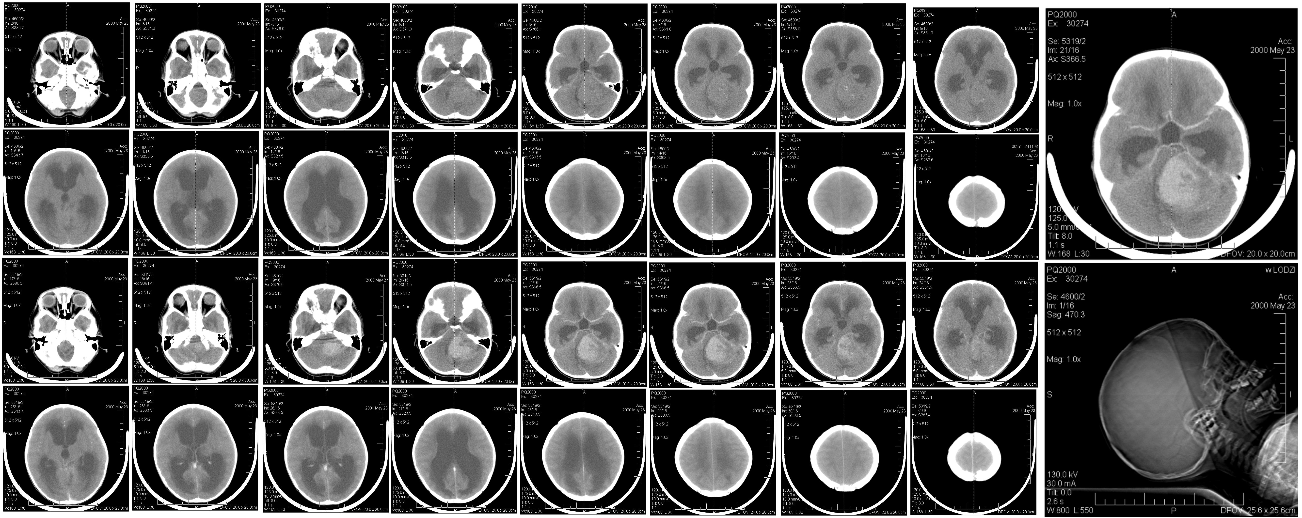
In children, roughly half of CNS tumours arise in the posterior fossa, with the remainder split between supratentorial and, far less commonly, spinal locations. Tumour type and biology vary strikingly by age, with embryonal tumours (medulloblastoma, ATRT, ETMR) clustering in early childhood and germ cell tumours and craniopharyngiomas more common in older children and adolescents.

References used here

  1. WHO Classification of Tumours Editorial Board. WHO Classification of Tumours: Central Nervous System Tumours. 5th Edition (Vol 6 of WHO Classification of Tumours series). IARC / WHO, 2021. ISBN: 978-92-832-4508-7.
  2. Louis DN, Perry A, Wesseling P, Brat DJ, Cree IA, Figarella-Branger D, Hawkins C, Ng HK, Pfister SM, Reifenberger G, Soffietti R, von Deimling A, Ellison DW. The 2021 WHO Classification of Tumors of the Central Nervous System: a summary. Neuro Oncol. 2021;23(8):1231-1251.
  3. Albright AL, Pollack IF, Adelson PD. Principles and Practice of Pediatric Neurosurgery. 3rd Edition. Thieme, 2015. ISBN: 978-1-60406-799-6.
  4. Winn HR (Editor). Youmans and Winn Neurological Surgery. 8th Edition (4-volume set). Elsevier, 2022. ISBN: 978-0-323-66192-8.
WHO Grade 4

Medulloblastoma

WHO 2021 classification: Embryonal tumour, CNS; Medulloblastoma (molecularly defined: WNT-activated; SHH-activated and TP53-wildtype; SHH-activated and TP53-mutant; non-WNT/non-SHH, Group 3 / Group 4).

The most common malignant pediatric brain tumour and the prototypical posterior-fossa embryonal tumour of childhood. WHO 2021 retains four molecular subgroups, each with distinct biology, prognosis, and emerging risk-adapted therapy.

Non-contrast CT in a 6-year-old child showing a posterior-fossa mass with obstructive hydrocephalus
Non-contrast CT brain in a 6-year-old girl with a hyperdense midline posterior-fossa mass (medulloblastoma) obstructing the fourth ventricle and producing supratentorial obstructive hydrocephalus. Hyperdensity on non-contrast CT reflects the high cellularity characteristic of embryonal tumours.
Credit: Reytan · License: Public domain · Source: Wikimedia Commons ↗

Epidemiology

Incidence
Approximately 20% of pediatric CNS tumours; most common malignant brain tumour in children.
Age peak
Bimodal in childhood; majority diagnosed between 3 and 8 years.
Location
Cerebellar vermis / fourth ventricle (children); cerebellar hemisphere more typical in adolescents and adults.

Clinical Presentation

  • Raised intracranial pressure from fourth-ventricular obstruction: morning headache, vomiting, lethargy, papilloedema.
  • Truncal and gait ataxia; nystagmus; head tilt with hydrocephalus.
  • Leptomeningeal dissemination at presentation in roughly one-third of patients; full-axis MRI and CSF cytology are mandatory before any adjuvant therapy.

Imaging

  • Hyperdense midline cerebellar mass on non-contrast CT (reflecting high cellularity).
  • MRI: T1 hypointense, T2 iso- to hypointense, restricted diffusion, heterogeneous enhancement.
  • Full neuraxis MRI and lumbar CSF for staging (Chang M-stage).
  • WNT tumours classically arise at the cerebellar peduncle / cerebellopontine angle; Group 3/4 tend to be midline.

Pathology & Molecular

Histology. Densely cellular small round blue cell tumour with high mitotic rate; Homer Wright rosettes may be present. Histological variants include classic, desmoplastic/nodular, extensive nodularity (MBEN), and large-cell / anaplastic, but molecular subgroup now drives risk-stratification.

Molecular. Four consensus molecular subgroups: WNT-activated (CTNNB1 mutation; favourable); SHH-activated (PTCH1/SMO/SUFU; further split by TP53 status; TP53-mutant SHH is a separate WHO entity with markedly worse outcome); non-WNT/non-SHH Group 3 (often MYC-driven, highest metastatic risk); non-WNT/non-SHH Group 4 (the largest subgroup overall).

Management

Surgery. Maximal safe resection of the primary tumour; degree of resection remains prognostic. Posterior-fossa syndrome (cerebellar mutism) is a recognised post-operative complication; counsel families pre-operatively.

Adjuvant therapy. Risk-adapted craniospinal radiotherapy (CSI) plus posterior-fossa boost, combined with multi-agent chemotherapy (vincristine, cisplatin, lomustine/CCNU and/or cyclophosphamide, the Packer-type regimen historically used in average-risk disease). Infants (<3 years) are typically treated with intensified chemotherapy and, where feasible, deferred or reduced-dose radiotherapy to mitigate neurocognitive toxicity.

Considerations. Subgroup-directed risk-stratification is now the basis of contemporary cooperative-group trials, with active investigation of de-escalation in WNT and chemotherapy-only regimens in infant SHH/MBEN disease.

Outcomes

5-year overall survival broadly 70-85% across pediatric medulloblastoma series in cooperative-group trials, but varies markedly by subgroup and risk category.

By molecular subgroup: WNT: excellent (>90% in standard-risk children). SHH, TP53-wildtype: intermediate. SHH, TP53-mutant: poor. Group 3, MYC-amplified / metastatic: poor. Group 4: intermediate, with progression often late.

Clinical Pearls

  • MRI of the whole neuraxis must precede surgical resection where feasible; post-operative blood products and dural enhancement complicate staging.
  • Molecular subgrouping (now routine in expert centres) changes therapy and prognostic counselling; histology alone is insufficient.
  • Cerebellar mutism / posterior fossa syndrome is more frequent with brainstem invasion and midline vermian resections.

Medulloblastoma molecular subgroups (WHO 2021)

SubgroupApprox. frequencyHallmark molecular featuresTypical agePrognosis (overall)
WNT-activated~10%CTNNB1 mutation; monosomy 6Older children; rare <4 yExcellent (5-y OS >90% in standard-risk)
SHH-activated, TP53-wildtype~15-20%PTCH1, SMO, SUFU; intact TP53Bimodal; infants and adolescentsIntermediate to good
SHH-activated, TP53-mutant~5%TP53 mutation (often germline; Li-Fraumeni)Older children / young adultsPoor
Non-WNT/non-SHH, Group 3~25%Frequent MYC amplification; metastasis commonInfants and young childrenPoor (worst in MYC-amplified)
Non-WNT/non-SHH, Group 4~35%Often chromosome 11 loss, 17q gain; isochromosome 17qChildren and adolescentsIntermediate
Adapted from the WHO Classification of CNS Tumours 5th Edition (2021) and Northcott et al. Nat Rev Dis Primers 2019.

References used here

  1. WHO Classification of Tumours Editorial Board. WHO Classification of Tumours: Central Nervous System Tumours. 5th Edition (Vol 6 of WHO Classification of Tumours series). IARC / WHO, 2021. ISBN: 978-92-832-4508-7.
  2. Louis DN, Perry A, Wesseling P, Brat DJ, Cree IA, Figarella-Branger D, Hawkins C, Ng HK, Pfister SM, Reifenberger G, Soffietti R, von Deimling A, Ellison DW. The 2021 WHO Classification of Tumors of the Central Nervous System: a summary. Neuro Oncol. 2021;23(8):1231-1251.
  3. Northcott PA, Robinson GW, Kratz CP, Mabbott DJ, Pomeroy SL, Clifford SC, Rutkowski S, Ellison DW, Malkin D, Taylor MD, Gajjar A, Pfister SM. Medulloblastoma. Nat Rev Dis Primers. 2019;5(1):11.
  4. Albright AL, Pollack IF, Adelson PD. Principles and Practice of Pediatric Neurosurgery. 3rd Edition. Thieme, 2015. ISBN: 978-1-60406-799-6.
  5. Winn HR (Editor). Youmans and Winn Neurological Surgery. 8th Edition (4-volume set). Elsevier, 2022. ISBN: 978-0-323-66192-8.
WHO Grade 1

Pilocytic Astrocytoma

WHO 2021 classification: Circumscribed astrocytic glioma; Pilocytic astrocytoma.

The most common pediatric brain tumour overall and the prototypical curable low-grade glioma. The vast majority are driven by a single MAPK-pathway alteration, most often the KIAA1549::BRAF fusion.

Histopathology of pilocytic astrocytoma, H&E stain at 200x magnification
Histopathological section of pilocytic astrocytoma (WHO Grade 1) at 200× magnification with haematoxylin and eosin staining, showing the biphasic pattern of compact piloid (hair-like) areas alternating with looser microcystic regions.
Credit: Marvin 101 · License: CC BY-SA 3.0 · Source: Wikimedia Commons ↗
Rosenthal fibers in pilocytic astrocytoma, H&E stain at 400x magnification
Rosenthal fibres; elongated, brightly eosinophilic, corkscrew-shaped structures within the cytoplasmic processes of neoplastic astrocytes. A characteristic (though not pathognomonic) histological feature of pilocytic astrocytoma. H&E stain, 400×.
Credit: Marvin 101 · License: CC BY-SA 3.0 · Source: Wikimedia Commons ↗

Epidemiology

Incidence
Roughly 15-20% of pediatric CNS tumours; the single most common pediatric primary brain tumour.
Age peak
First decade of life, with a slow tail into early adulthood.
Location
Cerebellum (most common), optic pathway/hypothalamus, brainstem (especially dorsal exophytic), supratentorial midline, and rarely spinal cord.

Clinical Presentation

  • Cerebellar lesions: ataxia, dysmetria, and signs of obstructive hydrocephalus.
  • Optic pathway lesions: progressive vision loss, proptosis, or, in NF1, often discovered on surveillance imaging.
  • Hypothalamic lesions: diencephalic syndrome in infants (failure to thrive despite normal caloric intake).
  • Brainstem lesions (dorsal exophytic): chronic cranial nerve palsies and gait disturbance.

Imaging

  • Classic cerebellar appearance: a well-circumscribed cyst with an avidly enhancing mural nodule.
  • Optic pathway: fusiform enlargement of nerve / chiasm; usually does not enhance dramatically.
  • Restricted diffusion is uncommon and would prompt reconsideration of grade.

Pathology & Molecular

Histology. Biphasic; compact piloid (hair-like) areas alternating with loose microcystic areas. Rosenthal fibres and eosinophilic granular bodies are characteristic. Microvascular proliferation can be present and does not, by itself, indicate higher grade.

Molecular. MAPK-pathway alteration in the great majority: KIAA1549::BRAF fusion (most common, especially in cerebellar tumours), BRAF V600E mutation in a smaller subset (often non-cerebellar), and NF1 loss in NF1-associated optic pathway gliomas. FGFR1 and other MAPK alterations occur uncommonly.

Management

Surgery. Gross-total resection is curative for accessible (e.g., cerebellar) tumours and is the goal where it can be achieved without unacceptable functional cost.

Adjuvant therapy. For unresectable, progressive, or symptomatic disease (especially optic pathway / hypothalamic): observation is often appropriate first-line, particularly in NF1. When treatment is required, vincristine/carboplatin chemotherapy is a long-standing standard; targeted therapy (MEK inhibitors such as selumetinib, and BRAF/MEK combinations for BRAF V600E disease) has become a major second-line option, supported by trial-level evidence.

Considerations. Radiotherapy is generally avoided in young children when alternative options exist, due to long-term neurocognitive, endocrine, and vascular morbidity.

Outcomes

10-year overall survival exceeds 90% for completely resected cerebellar pilocytic astrocytoma. Outcomes for unresectable midline tumours are more variable and depend on response to medical therapy and visual / endocrine sequelae.

Clinical Pearls

  • A cystic cerebellar mass with an enhancing mural nodule in a child is pilocytic astrocytoma until proven otherwise.
  • Treat the patient, not the scan; many NF1-associated optic pathway gliomas can be safely observed.
  • Confirm BRAF V600E before considering BRAF/MEK inhibitor therapy, as it predicts response.

MAPK-pathway alterations in pilocytic astrocytoma by location (typical distribution)

LocationMost common alterationOther alterations seen
CerebellarKIAA1549::BRAF fusion (majority)BRAF V600E (uncommon); other MAPK fusions
Optic pathway / hypothalamicNF1 loss (in NF1 patients); KIAA1549::BRAF (sporadic)FGFR1 alterations (rare)
Brainstem (dorsal exophytic)KIAA1549::BRAF fusionBRAF V600E
Supratentorial (non-midline)BRAF V600E or KIAA1549::BRAFFGFR1 alterations (rare)
Single MAPK-pathway alterations account for the great majority of cases; co-occurrence is unusual.

References used here

  1. WHO Classification of Tumours Editorial Board. WHO Classification of Tumours: Central Nervous System Tumours. 5th Edition (Vol 6 of WHO Classification of Tumours series). IARC / WHO, 2021. ISBN: 978-92-832-4508-7.
  2. Louis DN, Perry A, Wesseling P, Brat DJ, Cree IA, Figarella-Branger D, Hawkins C, Ng HK, Pfister SM, Reifenberger G, Soffietti R, von Deimling A, Ellison DW. The 2021 WHO Classification of Tumors of the Central Nervous System: a summary. Neuro Oncol. 2021;23(8):1231-1251.
  3. Fangusaro J, Onar-Thomas A, Young Poussaint T, Wu S, Ligon AH, Lindeman N, Banerjee A, Packer RJ, Kilburn LB, Goldman S, et al.. Selumetinib in paediatric patients with BRAF-aberrant or neurofibromatosis type 1-associated recurrent, refractory, or progressive low-grade glioma: a multicentre, phase 2 trial. Lancet Oncol. 2019;20(7):1011-1022.
  4. Albright AL, Pollack IF, Adelson PD. Principles and Practice of Pediatric Neurosurgery. 3rd Edition. Thieme, 2015. ISBN: 978-1-60406-799-6.
WHO Grade 4

Diffuse Midline Glioma, H3 K27-altered

WHO 2021 classification: Pediatric-type diffuse high-grade glioma; Diffuse midline glioma, H3 K27-altered.

The molecularly defined successor to the clinico-radiological term 'DIPG' (diffuse intrinsic pontine glioma). Defined by loss of H3 K27 trimethylation, most often due to a H3K27M mutation in H3F3A or HIST1H3B/C, or, less commonly, EZHIP overexpression.

Epidemiology

Incidence
Rare in absolute terms, but the dominant cause of pediatric brain-tumour mortality.
Age peak
Median age at diagnosis around 6-9 years; can occur in adolescents and rarely adults.
Location
Pons (classic 'DIPG' presentation), thalamus, and spinal cord; all midline structures.

Clinical Presentation

  • Pontine disease typically presents with a short prodrome (weeks) of the classic 'triad': multiple cranial neuropathies, long-tract signs, and cerebellar dysfunction.
  • Thalamic disease may present with hemiparesis, movement disorder, or hydrocephalus.
  • Endocrinopathy is uncommon; sudden personality change or severe headache should prompt urgent imaging.

Imaging

  • Pontine DMG: an expansile, T2-hyperintense pontine mass typically encasing the basilar artery; enhancement is variable and often minimal at diagnosis.
  • Diffuse infiltration without a clear cyst-and-nodule architecture distinguishes DMG from pilocytic astrocytoma and brainstem cavernoma.

Pathology & Molecular

Histology. Infiltrative astrocytic neoplasm with variable cellular atypia; classic high-grade features (mitoses, necrosis, microvascular proliferation) may or may not be present, and importantly, molecular criteria override histology, so a histologically lower-grade-appearing midline glioma with H3 K27 alteration is graded as WHO 4.

Molecular. H3 K27M mutation in H3F3A (H3.3) or HIST1H3B/C (H3.1); alternatively, EZHIP overexpression with retained wild-type H3 but loss of H3 K27 trimethylation. The unifying epigenetic consequence is loss of H3K27me3 and global dysregulation of polycomb repression.

Management

Surgery. Stereotactic biopsy is now the standard initial procedure in expert centres; it is feasible with acceptable safety and is essential for molecular diagnosis and trial enrollment. Maximal safe resection has a role in some non-pontine (e.g., thalamic, spinal) lesions.

Adjuvant therapy. Focal radiotherapy (typically 54 Gy in conventional fractions) remains the only intervention with reproducible symptomatic benefit and is the standard of care. No systemic therapy has yet demonstrated a survival advantage in randomised trials; clinical-trial enrollment is strongly encouraged.

Considerations. Re-irradiation at progression is increasingly offered and is associated with a temporary symptomatic and imaging response in a meaningful fraction of patients in published series.

Outcomes

Pontine DMG remains a uniformly fatal disease in contemporary series, with median overall survival of roughly 9-12 months from diagnosis. Non-pontine (thalamic, spinal) DMG may have somewhat longer survival but the disease is incurable.

Clinical Pearls

  • Stop saying 'DIPG' alone, the WHO 2021 entity is 'diffuse midline glioma, H3 K27-altered', and the diagnosis is molecular.
  • Biopsy is appropriate, ethical, and increasingly the standard at experienced centres; counselling should reflect this.
  • Symptom-directed steroid management and proactive palliative-care involvement are integral to high-quality care.

References used here

  1. WHO Classification of Tumours Editorial Board. WHO Classification of Tumours: Central Nervous System Tumours. 5th Edition (Vol 6 of WHO Classification of Tumours series). IARC / WHO, 2021. ISBN: 978-92-832-4508-7.
  2. Louis DN, Perry A, Wesseling P, Brat DJ, Cree IA, Figarella-Branger D, Hawkins C, Ng HK, Pfister SM, Reifenberger G, Soffietti R, von Deimling A, Ellison DW. The 2021 WHO Classification of Tumors of the Central Nervous System: a summary. Neuro Oncol. 2021;23(8):1231-1251.
  3. Winn HR (Editor). Youmans and Winn Neurological Surgery. 8th Edition (4-volume set). Elsevier, 2022. ISBN: 978-0-323-66192-8.
  4. Albright AL, Pollack IF, Adelson PD. Principles and Practice of Pediatric Neurosurgery. 3rd Edition. Thieme, 2015. ISBN: 978-1-60406-799-6.
WHO Grades 2-3 (and Subependymoma, Grade 1; Myxopapillary, Grade 2)

Ependymoma

WHO 2021 classification: Ependymal tumours, now anatomically- and molecularly-defined: Supratentorial ependymoma, ZFTA-fusion-positive (formerly RELA); Supratentorial ependymoma, YAP1-fusion-positive; Posterior-fossa ependymoma, group PFA; Posterior-fossa ependymoma, group PFB; Spinal ependymoma; Spinal ependymoma, MYCN-amplified; Myxopapillary ependymoma; Subependymoma.

A biologically heterogeneous family of tumours arising from ependymal lineage cells. The 2021 WHO classification, building on methylation-based work led by Pajtler and colleagues, defines ependymomas by anatomic compartment and molecular signature, with markedly different prognoses across groups.

Epidemiology

Incidence
Roughly 6-10% of pediatric CNS tumours.
Age peak
PFA infratentorial disease dominates in young children (median around 3 years); PFB occurs more in older children and adolescents; supratentorial ZFTA also tends to occur in younger children.
Location
Posterior fossa (most paediatric cases; floor of fourth ventricle, often extending through foramina of Luschka or Magendie); supratentorial intra-axial; or spinal (less common in children).

Clinical Presentation

  • Posterior-fossa: progressive hydrocephalus, ataxia, lower cranial neuropathies; subtle torticollis from cerebellar tonsillar herniation can be the only sign in infants.
  • Supratentorial: focal deficits and seizures depending on location.
  • Spinal: long-tract signs and back pain; myxopapillary tumours classically arise in the cauda equina/filum terminale.

Imaging

  • Posterior-fossa ependymoma classically extrudes through the foramina of Luschka or Magendie, helping distinguish it from medulloblastoma which more often fills the fourth ventricle without herniating out.
  • Heterogeneous enhancement with calcifications and small cysts is typical.
  • Whole neuraxis MRI is required given the risk of leptomeningeal dissemination, especially in PFA.

Pathology & Molecular

Histology. Perivascular pseudorosettes (consistent feature) and true ependymal rosettes (less common); GFAP-positive. Anaplastic features (mitoses, microvascular proliferation, necrosis) raise grade.

Molecular. Methylation-based grouping is now standard: PFA (CIMP-high, EZHIP-high, generally H3K27me3-low, worst prognosis); PFB (CIMP-low, better prognosis, often potentially curable with GTR alone in selected cases); ZFTA-fusion supratentorial ependymoma (formerly RELA, intermediate prognosis); YAP1-fusion (generally favourable); spinal MYCN-amplified (aggressive).

Management

Surgery. Gross-total resection is the single most important prognostic variable across all groups, the goal of every operation is maximal safe resection at first attempt. Second-look surgery is considered for residual disease before adjuvant therapy.

Adjuvant therapy. Adjuvant conformal radiotherapy is standard for WHO Grades 2-3 supratentorial and posterior-fossa ependymomas in children over 12-18 months. Chemotherapy has historically been used to defer radiotherapy in infants and to consolidate after GTR + RT, though its survival benefit in standard-risk disease is debated.

Considerations. Recurrence (most often local) is the main pattern of failure; salvage with re-resection ± re-irradiation may be appropriate.

Outcomes

Highly dependent on group and resection extent. PFB ependymoma after GTR has excellent long-term survival in published series. PFA ependymoma has substantially poorer 5-year survival, with progression-free survival often 50% or lower without aggressive multimodal therapy.

Clinical Pearls

  • Get a GTR the first time; it is the most modifiable variable affecting outcome.
  • A posterior-fossa mass extending out through the foramina of Luschka is ependymoma until proven otherwise.
  • Demand methylation profiling; PFA vs PFB fundamentally changes the conversation with the family.

Ependymoma molecular groups (WHO 2021 / Pajtler 2015 framework)

GroupLocationTypical ageKey molecular featurePrognosis
PFAPosterior fossaYoung children (median ~3 y)CIMP-high; EZHIP-high; low H3K27me3Poor (worst paediatric ependymoma group)
PFBPosterior fossaOlder children, adolescents, adultsCIMP-low; structural chromosomal aberrationsGood; can be curable with GTR alone in selected cases
ST-ZFTA (formerly RELA)SupratentorialChildren and young adultsZFTA fusion (most commonly ZFTA::RELA)Intermediate
ST-YAP1SupratentorialYoung childrenYAP1 fusion (e.g., YAP1::MAMLD1)Generally favourable
Spinal ependymomaSpinal cordAdolescents and adultsNF2 alterations commonFavourable with GTR
Spinal ependymoma, MYCN-amplifiedSpinal cord (especially cervical/thoracic)Young adultsMYCN amplificationAggressive; distinct WHO entity
MyxopapillaryConus medullaris / filum terminaleAdolescents and adultsDistinct epigenetic profileUsually favourable but recurrence with subtotal resection
Framework derives from Pajtler et al. Cancer Cell 2015 and is incorporated into the WHO Classification of CNS Tumours 5th Edition (2021).

References used here

  1. WHO Classification of Tumours Editorial Board. WHO Classification of Tumours: Central Nervous System Tumours. 5th Edition (Vol 6 of WHO Classification of Tumours series). IARC / WHO, 2021. ISBN: 978-92-832-4508-7.
  2. Louis DN, Perry A, Wesseling P, Brat DJ, Cree IA, Figarella-Branger D, Hawkins C, Ng HK, Pfister SM, Reifenberger G, Soffietti R, von Deimling A, Ellison DW. The 2021 WHO Classification of Tumors of the Central Nervous System: a summary. Neuro Oncol. 2021;23(8):1231-1251.
  3. Pajtler KW, Witt H, Sill M, Jones DTW, Hovestadt V, Kratochwil F, Wani K, Tatevossian R, Punchihewa C, Johann P, et al.. Molecular Classification of Ependymal Tumors across All CNS Compartments, Histopathological Grades, and Age Groups. Cancer Cell. 2015;27(5):728-743.
  4. Winn HR (Editor). Youmans and Winn Neurological Surgery. 8th Edition (4-volume set). Elsevier, 2022. ISBN: 978-0-323-66192-8.
WHO Grade 4

Atypical Teratoid/Rhabdoid Tumour (AT/RT)

WHO 2021 classification: Embryonal tumour, CNS; Atypical teratoid/rhabdoid tumour, defined by biallelic SMARCB1 (INI1) inactivation or, rarely, SMARCA4 (BRG1) inactivation.

A highly aggressive embryonal tumour of infancy and early childhood, biologically defined by loss of the SWI/SNF complex member SMARCB1 (most cases) or SMARCA4 (rare). Often confused histologically with medulloblastoma or PNET in older literature.

Epidemiology

Incidence
Rare overall but accounts for a disproportionate share of CNS tumours in children under 3 years.
Age peak
Most diagnosed before age 3; many before age 1.
Location
Approximately equally distributed between infratentorial (cerebellum, fourth ventricle, cerebellopontine angle) and supratentorial compartments; spinal involvement uncommon.

Clinical Presentation

  • In infants: lethargy, vomiting, head enlargement, regression of milestones, failure to thrive.
  • Cranial neuropathies and ataxia in cerebellopontine-angle disease.
  • Disseminated disease at presentation is common; full neuraxis imaging and CSF cytology required.

Imaging

  • Heterogeneous mass with frequent haemorrhage, necrosis, and calcification.
  • Restricted diffusion is typical, reflecting dense cellularity.
  • Often appears more aggressive on imaging than equivalent-sized medulloblastoma.

Pathology & Molecular

Histology. Pleomorphic small-blue-cell tumour with classical rhabdoid cells (eccentric nuclei, prominent nucleoli, eosinophilic inclusion-like cytoplasm). Loss of nuclear INI1 (SMARCB1) staining by immunohistochemistry is the diagnostic hallmark in the great majority of cases.

Molecular. Biallelic inactivation of SMARCB1 in over 95% of cases; a small minority show biallelic SMARCA4 loss with retained INI1 staining. Germline SMARCB1 (rhabdoid tumour predisposition syndrome) is identified in a substantial subset; genetic counselling and germline testing are mandatory.

Management

Surgery. Maximal safe resection is undertaken when feasible, but is often anatomically constrained.

Adjuvant therapy. Multimodal therapy combining intensive multi-agent chemotherapy ± high-dose chemotherapy with autologous stem-cell rescue, and radiotherapy (focal or craniospinal) where age permits. Outcomes have improved meaningfully with contemporary cooperative-group regimens.

Considerations. Genetic counselling and germline SMARCB1/SMARCA4 testing should be offered to every family at diagnosis.

Outcomes

Historically dismal; contemporary intensive regimens report 4-5 year overall survival broadly in the range of 30-50%, with selected favourable subgroups (older age, GTR, supratentorial location, no metastatic disease, ATRT-MYC or some ATRT-TYR molecular subtypes) doing better.

Clinical Pearls

  • Any rhabdoid-appearing or aggressive infant brain tumour requires SMARCB1 (INI1) immunohistochemistry; loss is diagnostic.
  • Germline testing belongs in the upfront workup, not as an afterthought.
  • Cooperative-group trial enrollment is the right default; outcomes outside specialised centres lag.

References used here

  1. WHO Classification of Tumours Editorial Board. WHO Classification of Tumours: Central Nervous System Tumours. 5th Edition (Vol 6 of WHO Classification of Tumours series). IARC / WHO, 2021. ISBN: 978-92-832-4508-7.
  2. Louis DN, Perry A, Wesseling P, Brat DJ, Cree IA, Figarella-Branger D, Hawkins C, Ng HK, Pfister SM, Reifenberger G, Soffietti R, von Deimling A, Ellison DW. The 2021 WHO Classification of Tumors of the Central Nervous System: a summary. Neuro Oncol. 2021;23(8):1231-1251.
  3. Winn HR (Editor). Youmans and Winn Neurological Surgery. 8th Edition (4-volume set). Elsevier, 2022. ISBN: 978-0-323-66192-8.
  4. Albright AL, Pollack IF, Adelson PD. Principles and Practice of Pediatric Neurosurgery. 3rd Edition. Thieme, 2015. ISBN: 978-1-60406-799-6.
WHO Grade 1

Craniopharyngioma

WHO 2021 classification: Tumours of the sellar region, Craniopharyngioma, adamantinomatous (adamantinomatous craniopharyngioma, ACP) and papillary (papillary craniopharyngioma, PCP), now classified as distinct entities reflecting their distinct molecular drivers.

A biologically benign but anatomically devastating tumour arising from remnants of Rathke's pouch in the sellar/suprasellar region. The 2021 WHO classification recognises adamantinomatous and papillary subtypes as separate entities, driven by CTNNB1 and BRAF V600E respectively.

Epidemiology

Incidence
Roughly 5-10% of pediatric brain tumours; the dominant non-glial supra-sellar mass in childhood.
Age peak
Bimodal: a paediatric peak (5-14 years, almost all adamantinomatous) and an adult peak (50-70 years, where papillary tumours predominate).
Location
Sellar and suprasellar; some tumours extend into the third ventricle or retrochiasmal cisterns.

Clinical Presentation

  • Endocrine: growth failure, hypothyroidism, central diabetes insipidus, delayed puberty, secondary adrenal insufficiency; endocrinopathy is often present at diagnosis and rarely fully recovers after surgery.
  • Visual: bitemporal hemianopia or asymmetric visual loss from chiasmal compression.
  • Mass effect: headache and hydrocephalus from third-ventricular obstruction.
  • Hypothalamic dysfunction with obesity and behavioural change may dominate the long-term clinical picture.

Imaging

  • Mixed solid/cystic suprasellar mass with calcification (especially in adamantinomatous tumours).
  • Cyst fluid characteristically described as 'machine-oil' at surgery in ACP.
  • Papillary tumours are typically solid, non-calcified, third-ventricular-floor masses in adults.

Pathology & Molecular

Histology. Adamantinomatous: epithelial nests with peripheral palisading, stellate reticulum, 'wet keratin' nodules. Papillary: well-differentiated squamous epithelium on fibrovascular cores.

Molecular. Adamantinomatous craniopharyngioma: somatic CTNNB1 (β-catenin) exon 3 mutations in the great majority. Papillary craniopharyngioma: BRAF V600E mutation in essentially all cases, a discovery that has opened the door to BRAF/MEK inhibitor therapy in selected adult patients.

Management

Surgery. The central trade-off is resection extent versus hypothalamic injury. Modern paediatric practice in expert centres favours a planned, anatomically tailored approach (transcranial, endoscopic endonasal, or combined) aiming for the maximum resection achievable without crossing into the hypothalamus. A planned subtotal resection followed by radiotherapy is increasingly accepted as a strategy to reduce hypothalamic morbidity.

Adjuvant therapy. Fractionated stereotactic radiotherapy or proton beam therapy is the standard adjuvant for residual or recurrent disease.

Considerations. Endocrine and ophthalmologic baseline assessment pre-operatively is mandatory. Long-term endocrine replacement (pituitary panel + ADH) and DI management often define quality of life.

Outcomes

10-year overall survival commonly exceeds 80-90% in paediatric series, but functional outcome (endocrine, visual, neurocognitive, hypothalamic-obesity) is variable and frequently impaired.

Clinical Pearls

  • Test for diabetes insipidus pre-operatively (paired plasma and urine osmolality, sodium, fluid balance); perioperative DI is the rule, not the exception.
  • The temptation to chase a 'GTR' across the hypothalamus is the most consequential decision in the operating room.
  • BRAF V600E status changes the medical-therapy conversation in recurrent papillary disease.

Adamantinomatous vs Papillary craniopharyngioma (WHO 2021)

FeatureAdamantinomatous (ACP)Papillary (PCP)
Age at presentationChildren (5-14 y peak); also adultsAdults (50-70 y peak); rare in childhood
Calcification on CTFrequent (>90%)Absent / rare
Cystic componentAlmost universal; 'machine-oil' fluidPredominantly solid
HistologyWet keratin, peripheral palisading, stellate reticulumMature squamous epithelium on fibrovascular cores
Molecular driverSomatic CTNNB1 (β-catenin) exon 3 mutationBRAF V600E in essentially all cases
Targeted therapyNone establishedBRAF/MEK inhibitors (selected adult cases)
Typical anatomic seatSellar and suprasellarThird-ventricle floor / suprasellar
WHO 2021 recognises ACP and PCP as distinct entities, reflecting their distinct molecular drivers identified by Brastianos et al. (Nat Genet 2014).

References used here

  1. WHO Classification of Tumours Editorial Board. WHO Classification of Tumours: Central Nervous System Tumours. 5th Edition (Vol 6 of WHO Classification of Tumours series). IARC / WHO, 2021. ISBN: 978-92-832-4508-7.
  2. Louis DN, Perry A, Wesseling P, Brat DJ, Cree IA, Figarella-Branger D, Hawkins C, Ng HK, Pfister SM, Reifenberger G, Soffietti R, von Deimling A, Ellison DW. The 2021 WHO Classification of Tumors of the Central Nervous System: a summary. Neuro Oncol. 2021;23(8):1231-1251.
  3. Brastianos PK, Taylor-Weiner A, Manley PE, Jones RT, Dias-Santagata D, Thorner AR, Lawrence MS, Rodriguez FJ, Bernardo LA, Schubert L, et al.. Exome sequencing identifies BRAF mutations in papillary craniopharyngiomas. Nat Genet. 2014;46(2):161-165.
  4. Albright AL, Pollack IF, Adelson PD. Principles and Practice of Pediatric Neurosurgery. 3rd Edition. Thieme, 2015. ISBN: 978-1-60406-799-6.
  5. Winn HR (Editor). Youmans and Winn Neurological Surgery. 8th Edition (4-volume set). Elsevier, 2022. ISBN: 978-0-323-66192-8.
Typically WHO Grade 1 (pilocytic astrocytoma)

Optic Pathway Glioma

WHO 2021 classification: Most are circumscribed astrocytic gliomas, predominantly pilocytic astrocytoma; rarer high-grade variants exist.

A distinct clinico-anatomic entity rather than a separate WHO tumour type. Approximately half of paediatric optic pathway gliomas (OPGs) arise in the setting of neurofibromatosis type 1, where they often behave indolently.

Epidemiology

Incidence
Approximately 5% of paediatric brain tumours; lifetime risk of OPG in NF1 is about 15-20%, of which a smaller fraction become symptomatic.
Age peak
First decade, especially under age 6.
Location
Optic nerves, chiasm, optic tracts, and posterior optic pathway / hypothalamus.

Clinical Presentation

  • Visual loss (often slowly progressive and difficult to elicit in young children), the most clinically meaningful endpoint.
  • Proptosis with isolated optic-nerve involvement.
  • Hypothalamic involvement: precocious puberty, diencephalic syndrome (infants), or hypopituitarism.
  • In NF1: many are discovered incidentally on routine surveillance imaging and never become symptomatic.

Imaging

  • Fusiform enlargement of one or both optic nerves, with or without tortuosity, in isolated optic-nerve disease.
  • Chiasmal/hypothalamic OPGs may be large and partly cystic; surrounding signal change in optic radiations is common in NF1.
  • Sporadic (non-NF1) OPGs tend to be more aggressive radiographically and clinically.

Pathology & Molecular

Histology. Most are pilocytic astrocytoma; pilomyxoid variants (more common in infants) traditionally were grade 2 but the 2021 WHO no longer assigns a grade to pilomyxoid astrocytoma as a separate entity, recognising its behaviour overlaps with pilocytic.

Molecular. NF1-associated OPGs are driven by biallelic NF1 loss in tumour cells. Sporadic OPGs more frequently harbour KIAA1549::BRAF fusions or BRAF V600E.

Management

Surgery. Biopsy is generally avoided in NF1-associated OPG when imaging is characteristic; biopsy may be appropriate in sporadic disease or atypical lesions. Debulking has a limited role and is reserved for selected symptomatic cases.

Adjuvant therapy. Observation is appropriate first-line in many NF1 patients. When treatment is required (progressive visual loss or radiological progression), vincristine/carboplatin remains a long-standing first-line chemotherapy. MEK inhibitors (selumetinib in particular) have demonstrated objective tumour response in paediatric NF1-associated and BRAF-aberrant low-grade gliomas in prospective trials, and have become an important option in this setting.

Considerations. Radiotherapy is generally avoided in young NF1 patients owing to risks of second malignancy, moyamoya, and neurocognitive injury.

Outcomes

Long-term overall survival is excellent (>90% at 10 years) for the great majority of NF1-associated OPGs. Visual outcomes are more variable and depend on tumour location, age at presentation, and response to therapy.

Clinical Pearls

  • Vision, not tumour size, is the primary clinical endpoint; pair every imaging follow-up with formal ophthalmology and visual-acuity testing appropriate to age.
  • Selumetinib and other MEK inhibitors have prospective trial data supporting use in BRAF-aberrant or NF1-associated paediatric low-grade glioma.
  • Counsel NF1 families that many OPGs never need treatment.

References used here

  1. WHO Classification of Tumours Editorial Board. WHO Classification of Tumours: Central Nervous System Tumours. 5th Edition (Vol 6 of WHO Classification of Tumours series). IARC / WHO, 2021. ISBN: 978-92-832-4508-7.
  2. Louis DN, Perry A, Wesseling P, Brat DJ, Cree IA, Figarella-Branger D, Hawkins C, Ng HK, Pfister SM, Reifenberger G, Soffietti R, von Deimling A, Ellison DW. The 2021 WHO Classification of Tumors of the Central Nervous System: a summary. Neuro Oncol. 2021;23(8):1231-1251.
  3. Fangusaro J, Onar-Thomas A, Young Poussaint T, Wu S, Ligon AH, Lindeman N, Banerjee A, Packer RJ, Kilburn LB, Goldman S, et al.. Selumetinib in paediatric patients with BRAF-aberrant or neurofibromatosis type 1-associated recurrent, refractory, or progressive low-grade glioma: a multicentre, phase 2 trial. Lancet Oncol. 2019;20(7):1011-1022.
  4. Dombi E, Baldwin A, Marcus LJ, Fisher MJ, Weiss B, Kim A, Whitcomb P, Martin S, Aschbacher-Smith LE, Rizvi TA, et al.. Activity of Selumetinib in Neurofibromatosis Type 1-Related Plexiform Neurofibromas. N Engl J Med. 2016;375(26):2550-2560.
  5. Albright AL, Pollack IF, Adelson PD. Principles and Practice of Pediatric Neurosurgery. 3rd Edition. Thieme, 2015. ISBN: 978-1-60406-799-6.
WHO Grade 4 (in most cases)

Pediatric-type Diffuse High-Grade Glioma (non-DMG)

WHO 2021 classification: Pediatric-type diffuse high-grade glioma; includes Diffuse hemispheric glioma, H3 G34-mutant; Diffuse pediatric-type high-grade glioma, H3-wildtype and IDH-wildtype; and Infant-type hemispheric glioma.

WHO 2021 separates paediatric high-grade gliomas from their adult counterparts based on distinct molecular drivers. They are biologically and clinically different diseases from adult IDH-wildtype glioblastoma.

Epidemiology

Incidence
Rare overall; together with diffuse midline glioma, account for the bulk of paediatric high-grade glioma diagnoses.
Age peak
Variable by entity; infant-type hemispheric glioma in infancy; H3 G34-mutant disease in adolescents and young adults.
Location
Cerebral hemispheres (often parietal/temporal for H3 G34 disease).

Clinical Presentation

  • Seizures, focal deficits, and signs of raised ICP; duration of prodrome is shorter than for low-grade glioma.
  • Infant-type hemispheric glioma may present with macrocephaly, large heterogeneous hemispheric mass, and surprisingly long survival despite high-grade histology.

Imaging

  • Hemispheric mass with enhancement, surrounding T2 hyperintensity, and frequent necrosis.
  • Multifocal disease is uncommon at presentation.

Pathology & Molecular

Histology. Diffusely infiltrating astrocytic neoplasm with mitoses, microvascular proliferation, and/or necrosis (one or both for WHO Grade 4 designation by historical histological criteria).

Molecular. H3 G34-mutant: somatic H3F3A G34R/V mutation, almost always co-occurring with TP53 and ATRX alterations. Infant-type hemispheric glioma: receptor-tyrosine-kinase fusions, classically involving NTRK family, ROS1, ALK, or MET; opening the door to targeted therapy.

Management

Surgery. Maximal safe resection is the foundation.

Adjuvant therapy. Focal radiotherapy is standard in children old enough to tolerate it; chemotherapy choice (temozolomide and other regimens) has been extrapolated from adult glioblastoma trials, with cooperative-group trial enrollment encouraged. Infant-type hemispheric glioma with a defined RTK fusion is a candidate for targeted therapy (e.g., TRK inhibitors for NTRK fusions) under appropriate supervision.

Considerations. Comprehensive molecular profiling at diagnosis is essential, both for accurate WHO classification and for identification of actionable targets.

Outcomes

H3 G34-mutant glioma: median overall survival historically 18-24 months in adolescents/young adults. Infant-type hemispheric glioma: survival is notably better than histology alone would suggest, particularly when a targetable RTK fusion is present.

Clinical Pearls

  • Histology alone is no longer sufficient; every paediatric high-grade glioma needs molecular profiling at diagnosis.
  • An infant with a 'glioblastoma-like' hemispheric mass is a candidate for fusion profiling and possibly targeted therapy.

References used here

  1. WHO Classification of Tumours Editorial Board. WHO Classification of Tumours: Central Nervous System Tumours. 5th Edition (Vol 6 of WHO Classification of Tumours series). IARC / WHO, 2021. ISBN: 978-92-832-4508-7.
  2. Louis DN, Perry A, Wesseling P, Brat DJ, Cree IA, Figarella-Branger D, Hawkins C, Ng HK, Pfister SM, Reifenberger G, Soffietti R, von Deimling A, Ellison DW. The 2021 WHO Classification of Tumors of the Central Nervous System: a summary. Neuro Oncol. 2021;23(8):1231-1251.
  3. Winn HR (Editor). Youmans and Winn Neurological Surgery. 8th Edition (4-volume set). Elsevier, 2022. ISBN: 978-0-323-66192-8.
Papilloma (WHO Grade 1), Atypical papilloma (Grade 2), Carcinoma (Grade 3)

Choroid Plexus Tumours

WHO 2021 classification: Choroid plexus tumours; Choroid plexus papilloma, Atypical choroid plexus papilloma, Choroid plexus carcinoma.

A spectrum of tumours arising from choroid plexus epithelium, predominantly in infants and young children. Choroid plexus carcinoma carries a strong association with germline TP53 mutations (Li-Fraumeni syndrome).

Epidemiology

Incidence
Roughly 2-5% of paediatric brain tumours overall, but substantially over-represented in infants (a leading cause of intraventricular mass in the first year of life).
Age peak
First two years for most; carcinomas particularly cluster in infancy.
Location
Lateral ventricle (most common in children), fourth ventricle, third ventricle, or cerebellopontine angle.

Clinical Presentation

  • Hydrocephalus from CSF overproduction and/or obstruction is the dominant presenting feature in infants; head enlargement, bulging fontanelle, vomiting.
  • Older children may present with focal deficit, seizures, or raised ICP.

Imaging

  • Avidly enhancing intraventricular mass, often with a cauliflower-like lobulated surface.
  • Hydrocephalus is essentially universal at diagnosis.
  • Papillomas tend to be well-circumscribed; carcinomas show necrosis, parenchymal invasion, and oedema.

Pathology & Molecular

Histology. Papilloma: papillary architecture with bland epithelium resembling normal choroid plexus. Atypical: increased mitoses, hypercellularity, blurred papillary architecture. Carcinoma: frank malignant features; high mitotic rate, necrosis, nuclear pleomorphism, brain invasion.

Molecular. Choroid plexus carcinoma is strongly associated with germline TP53 mutations (Li-Fraumeni syndrome) and germline testing is part of standard workup.

Management

Surgery. Gross-total resection is the cornerstone of management for all grades; vascular control of choroidal arteries is the key technical challenge given the highly vascular nature of these tumours and the typical infantile patient.

Adjuvant therapy. Papilloma: GTR alone is usually curative. Carcinoma: post-operative chemotherapy is standard, with radiotherapy considered in older children where age-appropriate. Treatment of recurrence is individualised.

Considerations. Hydrocephalus may resolve after tumour resection, but a substantial fraction of patients still require a permanent CSF-diversion procedure.

Outcomes

Papilloma: long-term overall survival approaches 100% after GTR. Atypical papilloma: intermediate. Carcinoma: 5-year overall survival in the broad range of 40-60% in modern series, heavily dependent on resection extent and germline TP53 status.

Clinical Pearls

  • Pre-operative embolisation can be considered for large, hypervascular paediatric choroid plexus tumours where feasible.
  • Always offer germline TP53 testing in choroid plexus carcinoma, the implications for family screening are substantial.
  • Do not assume hydrocephalus will resolve after tumour resection; plan for follow-up imaging and clinical surveillance.

References used here

  1. WHO Classification of Tumours Editorial Board. WHO Classification of Tumours: Central Nervous System Tumours. 5th Edition (Vol 6 of WHO Classification of Tumours series). IARC / WHO, 2021. ISBN: 978-92-832-4508-7.
  2. Louis DN, Perry A, Wesseling P, Brat DJ, Cree IA, Figarella-Branger D, Hawkins C, Ng HK, Pfister SM, Reifenberger G, Soffietti R, von Deimling A, Ellison DW. The 2021 WHO Classification of Tumors of the Central Nervous System: a summary. Neuro Oncol. 2021;23(8):1231-1251.
  3. Albright AL, Pollack IF, Adelson PD. Principles and Practice of Pediatric Neurosurgery. 3rd Edition. Thieme, 2015. ISBN: 978-1-60406-799-6.
  4. Winn HR (Editor). Youmans and Winn Neurological Surgery. 8th Edition (4-volume set). Elsevier, 2022. ISBN: 978-0-323-66192-8.
Variable by histology

CNS Germ Cell Tumours

WHO 2021 classification: Germ cell tumours of the CNS; Germinoma; Embryonal carcinoma; Yolk sac tumour; Choriocarcinoma; Teratoma (mature, immature, with malignant transformation); Mixed germ cell tumours.

An anatomically restricted but clinically distinct group of midline tumours, classically pineal or suprasellar, with a strong adolescent male predominance for pineal disease. Pure germinoma is exquisitely radio- and chemo-sensitive; non-germinomatous germ cell tumours (NGGCT) are more aggressive but still potentially curable.

Epidemiology

Incidence
Roughly 3-5% of paediatric CNS tumours in Western series; substantially higher in East Asian populations.
Age peak
Adolescence; pineal disease has a strong male predominance, suprasellar disease less so.
Location
Pineal region (most common, male predominance), suprasellar (similar sex distribution), and rarely both ('bifocal' disease, considered germinoma until proven otherwise).

Clinical Presentation

  • Pineal: Parinaud syndrome (upgaze palsy, light-near dissociation, convergence-retraction nystagmus) and obstructive hydrocephalus.
  • Suprasellar: central diabetes insipidus (often the earliest and most specific symptom), visual loss, hypopituitarism, and growth failure; DI without a clearly visible suprasellar mass should prompt repeat imaging and pituitary stalk assessment.
  • Bifocal: features of both.

Imaging

  • Pineal mass with hydrocephalus; calcification is often present (germ cells typically engulf the pineal calcification).
  • Suprasellar: thickening of the pituitary stalk or a suprasellar mass.
  • Whole-neuraxis MRI plus CSF cytology are standard staging; serum and CSF tumour markers (β-hCG, α-fetoprotein) are essential.

Pathology & Molecular

Histology. Germinoma resembles testicular seminoma; uniform large cells with a lymphocytic infiltrate. NGGCTs include teratoma (mature: cystic, with skin/cartilage/teeth; immature: with embryonic elements), yolk sac tumour (Schiller-Duval bodies, AFP-positive), embryonal carcinoma, choriocarcinoma (β-hCG-secreting), and mixed forms.

Molecular. KIT/RAS-pathway alterations described in pure germinoma; teratomatous and NGGCT components have more complex genomes.

Management

Surgery. Pure germinoma with positive markers does not necessarily require resection; tissue diagnosis (endoscopic biopsy or stereotactic) plus CSF diversion (often endoscopic third ventriculostomy through the same approach for pineal disease) is the standard. Mature teratoma should be resected. NGGCT often requires planned 'second-look' surgery after neoadjuvant chemotherapy to address residual non-germinomatous components.

Adjuvant therapy. Germinoma: combined chemotherapy (carboplatin/etoposide-based) and reduced-volume / reduced-dose radiotherapy (commonly whole-ventricular RT for localised disease, with craniospinal RT for metastatic disease) is highly effective. NGGCT: intensive multi-agent chemotherapy plus focal or craniospinal RT.

Considerations. Endocrine follow-up is long-term and central to quality of life, particularly for suprasellar germ cell tumours.

Outcomes

Pure intracranial germinoma: 10-year overall survival commonly >90%. NGGCT: 5-year overall survival generally in the 70-80% range with contemporary regimens, lower for poor-prognosis subtypes.

Clinical Pearls

  • Always measure serum and CSF AFP and β-hCG before any surgery; they change the diagnosis, the prognosis, and the surgical plan.
  • Persistent unexplained central diabetes insipidus in a child mandates dedicated MRI of the pituitary stalk and follow-up imaging; germinoma may be invisible at first.
  • An ETV through the same endoscopic approach as a pineal biopsy treats both the hydrocephalus and the diagnostic question in one stage.

CNS germ cell tumours; markers and clinical implications

TumourSerum/CSF AFPSerum/CSF β-hCGSurgical roleSensitivity to chemotherapy & radiation
Pure germinomaNormalNormal or mildly elevated (syncytiotrophoblast cells)Biopsy only; no resection requiredVery high; combined chemo + reduced-volume RT highly curative
Mature teratomaNormalNormalResection (chemo/RT not effective)Resistant; surgery is the treatment
Immature teratomaMay be mildly elevatedNormalResection ± chemo for residualIntermediate
Yolk sac tumourMarkedly elevatedNormalResection of residual after chemoModerate; needs intensive chemo + RT
ChoriocarcinomaNormalMarkedly elevatedResection of residual after chemoModerate
Embryonal carcinomaVariableVariableResection of residual after chemoModerate
Mixed NGGCTVariableVariablePlanned second-look resection after neoadjuvant chemoVariable; protocol-driven multimodal therapy
Marker elevation (in either serum or CSF) is essentially diagnostic of NGGCT components and changes the treatment plan; markers should always be drawn pre-operatively.

References used here

  1. WHO Classification of Tumours Editorial Board. WHO Classification of Tumours: Central Nervous System Tumours. 5th Edition (Vol 6 of WHO Classification of Tumours series). IARC / WHO, 2021. ISBN: 978-92-832-4508-7.
  2. Louis DN, Perry A, Wesseling P, Brat DJ, Cree IA, Figarella-Branger D, Hawkins C, Ng HK, Pfister SM, Reifenberger G, Soffietti R, von Deimling A, Ellison DW. The 2021 WHO Classification of Tumors of the Central Nervous System: a summary. Neuro Oncol. 2021;23(8):1231-1251.
  3. Albright AL, Pollack IF, Adelson PD. Principles and Practice of Pediatric Neurosurgery. 3rd Edition. Thieme, 2015. ISBN: 978-1-60406-799-6.
  4. Winn HR (Editor). Youmans and Winn Neurological Surgery. 8th Edition (4-volume set). Elsevier, 2022. ISBN: 978-0-323-66192-8.
WHO Grade 4

Embryonal Tumour with Multilayered Rosettes (ETMR)

WHO 2021 classification: Embryonal tumours, CNS; Embryonal tumour with multilayered rosettes (ETMR), defined by amplification or fusion of the C19MC microRNA cluster on chromosome 19q13.42, or, in rare cases, by DICER1 alterations.

A relatively recently codified entity unifying lesions previously described as ETANTR (embryonal tumour with abundant neuropil and true rosettes), ependymoblastoma, and medulloepithelioma. Defined molecularly by C19MC alteration in the great majority.

Epidemiology

Incidence
Rare in absolute terms.
Age peak
Under 4 years; most cases in infants and toddlers.
Location
Most often supratentorial (cerebral hemispheres), but can arise in posterior fossa or brainstem.

Clinical Presentation

  • Rapid clinical progression with raised ICP, focal deficit, or seizures.
  • Often a large mass on first presentation, given the very young age and short prodrome.

Imaging

  • Large heterogeneous mass with necrosis, calcification, and restricted diffusion.
  • Often striking mass effect relative to symptom duration.

Pathology & Molecular

Histology. Small primitive cells, abundant neuropil, and characteristic multilayered (true) ependymoblastic-type rosettes.

Molecular. Amplification or fusion of the C19MC miRNA cluster (TTYH1::C19MC fusion is the most common). Rarely, biallelic DICER1 alteration defines a separate subset with overlapping morphology but distinct biology.

Management

Surgery. Maximal safe resection, often constrained by the large size of the tumour at presentation in a small child.

Adjuvant therapy. Intensive multi-agent chemotherapy ± high-dose chemotherapy with autologous stem-cell rescue; radiotherapy is challenging in the very young and is deferred or substituted with chemotherapy where possible.

Considerations. Cooperative-group trial enrollment is the appropriate default for this rare and aggressive disease.

Outcomes

Outcomes historically poor; modern intensive regimens have improved survival in selected patients but disease remains aggressive and a substantial proportion succumb to recurrence.

Clinical Pearls

  • C19MC amplification (FISH or methylation profiling) is the diagnostic anchor.
  • Treatment is most appropriately delivered within a high-volume paediatric neuro-oncology team and ideally on a cooperative-group trial.

References used here

  1. WHO Classification of Tumours Editorial Board. WHO Classification of Tumours: Central Nervous System Tumours. 5th Edition (Vol 6 of WHO Classification of Tumours series). IARC / WHO, 2021. ISBN: 978-92-832-4508-7.
  2. Louis DN, Perry A, Wesseling P, Brat DJ, Cree IA, Figarella-Branger D, Hawkins C, Ng HK, Pfister SM, Reifenberger G, Soffietti R, von Deimling A, Ellison DW. The 2021 WHO Classification of Tumors of the Central Nervous System: a summary. Neuro Oncol. 2021;23(8):1231-1251.
  3. Winn HR (Editor). Youmans and Winn Neurological Surgery. 8th Edition (4-volume set). Elsevier, 2022. ISBN: 978-0-323-66192-8.
Variable by histology

Pediatric Spinal Cord Tumours (Intramedullary)

WHO 2021 classification: Most paediatric intramedullary tumours are gliomas, most commonly pilocytic astrocytoma (WHO Grade 1) or ependymoma, or rarely high-grade gliomas, hemangioblastomas, or gangliogliomas.

Paediatric intramedullary spinal cord tumours are far less common than their intracranial counterparts. The differential differs from adults: paediatric intramedullary disease is dominated by astrocytic gliomas, whereas adult intramedullary disease is dominated by ependymoma.

Epidemiology

Incidence
Approximately 4-10% of paediatric CNS tumours.
Age peak
Throughout childhood; presentation often delayed.
Location
Cervical or upper thoracic cord more common than lumbar; long-segment involvement is typical.

Clinical Presentation

  • Insidious; back pain, gait change, focal weakness, scoliosis (a not-uncommon initial presentation), and sphincter disturbance.
  • The interval from first symptom to diagnosis is often months to years.

Imaging

  • Whole-spine MRI with contrast is essential; tumours can be long-segment with associated cysts ('cyst caps' above and below the solid tumour).
  • Astrocytoma: typically more diffuse, eccentric, less well-circumscribed than ependymoma.
  • Ependymoma: more central, well-circumscribed, often with haemorrhagic 'cap' sign.

Pathology & Molecular

Histology. Most paediatric intramedullary tumours are pilocytic astrocytoma or ependymoma; ganglioglioma, hemangioblastoma (often VHL-associated), and rarer high-grade gliomas (including spinal H3 K27-altered DMG and spinal MYCN-amplified ependymoma) make up the remainder.

Molecular. As per the relevant entity; KIAA1549::BRAF or BRAF V600E for pilocytic astrocytoma; molecular subgrouping for ependymoma; H3 K27 alteration for spinal DMG.

Management

Surgery. Maximal safe resection guided by intra-operative neurophysiological monitoring (motor and somatosensory evoked potentials, D-wave); ultrasound and where available intra-operative MRI assist in confirming extent.

Adjuvant therapy. Pilocytic astrocytoma: GTR alone often suffices. Ependymoma: GTR + selective adjuvant RT for residual or higher-grade disease. High-grade gliomas: focal RT ± systemic therapy.

Considerations. Spinal stability and risk of post-laminectomy or post-laminoplasty kyphosis must be considered, particularly in younger children; laminoplasty is generally preferred over laminectomy for multilevel exposure.

Outcomes

Pilocytic spinal cord astrocytoma after GTR: long-term overall survival exceeds 80-90% in published paediatric series. Ependymoma: similar with GTR. High-grade gliomas including spinal DMG: poor.

Clinical Pearls

  • Idiopathic scoliosis in a child with a non-classical curve or neurological signs warrants whole-spine MRI before brace or surgical treatment.
  • Intra-operative neuromonitoring is the standard of care for intramedullary tumour resection.
  • Laminoplasty rather than laminectomy reduces but does not eliminate the risk of post-operative spinal deformity.

References used here

  1. WHO Classification of Tumours Editorial Board. WHO Classification of Tumours: Central Nervous System Tumours. 5th Edition (Vol 6 of WHO Classification of Tumours series). IARC / WHO, 2021. ISBN: 978-92-832-4508-7.
  2. Louis DN, Perry A, Wesseling P, Brat DJ, Cree IA, Figarella-Branger D, Hawkins C, Ng HK, Pfister SM, Reifenberger G, Soffietti R, von Deimling A, Ellison DW. The 2021 WHO Classification of Tumors of the Central Nervous System: a summary. Neuro Oncol. 2021;23(8):1231-1251.
  3. Albright AL, Pollack IF, Adelson PD. Principles and Practice of Pediatric Neurosurgery. 3rd Edition. Thieme, 2015. ISBN: 978-1-60406-799-6.
  4. Winn HR (Editor). Youmans and Winn Neurological Surgery. 8th Edition (4-volume set). Elsevier, 2022. ISBN: 978-0-323-66192-8.